SCRIPTOR
Of all the papers and photographs produced in the Journal of the Biological Photographic Association and Journal of Biological Photography, I do not believe any have been so well known and so informative as the paper entitled, "Fluorescein of the Brain - The Photographic Procedure" by C. P. Hodge et al. This article appeared in JBPA , 1978, 46 (2), pps 67-79.
This work was presented to the Royal College of Surgeons, and the Royal College of Obstetrics and Gynecology of Great Britain, and received their Gold Medal Award. The work also helped secure an Honorary Fellowship of the Royal Photographic Society of Great Britain for the author. It is well worth a re-read, as its principles aptly apply to present-day digital recording.
Having said that, let us not let this splendid paper overshadow a very nice, concise, paper entitled "Monitoring Tar Therapeutics with Fluorescence Photography" by John Hendrix, JBPA , 1978, 46 (2), pps 80-81.
Finally, the book review of Jerome P. O'Neill on "The Collection, Use, and Care of Historical Photographs" authored by Robert A. Weinstein and Larry Booth, pps 55-56, is a valuable addition to any illustrator's library. It is easy and interesting to read, and contains an abundance of information on a topic that we should all be concerned about - How long will our illustrations last?
Scriptor
With the passing of Peter Hansell, Ron Irvine picks up
the pen to continue "25 Years ago in the Journal of Biological
Photography" and writes under the pseudonym SCRIPTOR. Ron is a
long-time member of BCA and IMI, is a Registered Biological Photographer,
Fellow of the BCA and an Honorary Fellow of IMI.
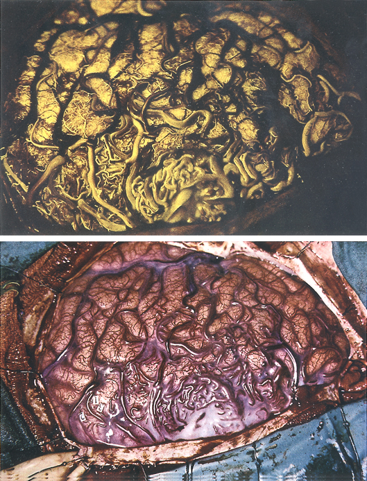
(Top) Fluorescein angiography in the arterial and capillary phase displays the circulatory supply of the angioma and provides the surgeon with precise detail for surgical removal. Thus, vital regions of the surface of the brain concerned with motor control and speech are avoided. Both photographs are of the left fronto-temporal region of the brain.
(Bottom) Color photograph of an abnormal collection of blood vessels (angioma)
on the surface of a brain exposed during surgery. It is impossible for the surgeon
to differentiate between arteries and abnormal red veins.