 |
||||
Nancy Grahame Joy: Envisioning Our Future |
Jennifer A. Polk and Nancy Grahame Joy was an influential leader in shaping medical illustration as an academic discipline in Canada, as chair of Art as Applied to Medicine (AAM) at the University of Toronto (U of T). Her approach to visual communication enhanced the standards of academic teaching material, and reflected the influence of her long professional relationship with anatomist John Charles Boileau Grant. Medical artists should be “born teachers, artists by vocation and scientists by nature.”
|
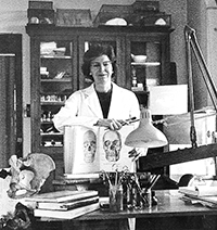
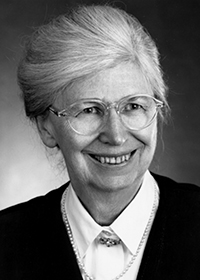
Origins Nancy Jean Hannah Grahame Joy was born in Toronto in 1920, the daughter of Ernst Grahame and Dorothy Ewart Joy (Canadian Who’s Who, 1983). Her father, a Toronto lawyer, had been a fighter pilot during WW1 (National Archives). Her mother had studied art with the Canadian artist Mary Wrinch Reid (Allen 1983). Her maternal grandfather, Alexander Primrose, was a surgeon, anatomist and, from 1920 to 1932, Dean of the Faculty of Medicine at the University of Toronto. These strands of intrepidity, art and science would figure in Nancy Joy’s subsequent career as a medical artist and as Chair of Art as Applied to Medicine (AAM) at the University of Toronto (U of T) (Figures 1 and 2). Education and early work From the age of 13, Joy knew she would become a medical illustrator (Joy, pers. comm., 2001). In 1939, she enrolled at the Ontario College of Art (OCA) in Toronto, where she completed a four-year diploma in drawing and painting. She had originally hoped to study medical art under Max Brödel at Johns Hopkins University after college, but Brödel retired in 1940, when Joy was only midway through her art studies. Instead, in 1942, she obtained special permission from Dr. J. C. B. Grant, Chair of Anatomy at the University of Toronto (U of T), to attend classes in anatomy, histology, embryology, and neuroanatomy with U of T’s medical students. During this time, Grant commissioned Joy to rework some of the drawings in his recently completed book A Method of Anatomy (1937) and, along with Dorothy Foster Chubb, to work as a freelance illustrator on the second volume of his first edition of An Atlas of Anatomy (1943). This venture with Grant was the beginning of what would turn out to be the longest working relationship of Joy’s career, spanning more than 30 years. Still seeking education in medical illustration, Joy was encouraged by Maria Torrance Wishart, director of the Department of Medical Art Service at U of T, to study under Tom Jones in the Department of Medical and Dental Illustration, University of Illinois at Chicago (UIC). In 1944 Joy registered in the Illinois program, which at that time was “very informal,” as she recalled in a 1973 interview (U of T Archives); after two months as a student, Joy was hired by Jones to work on his staff in the Illustration Studios at UIC. There she began to experiment with the construction of teaching models in plastics and other materials, and assisted in building exhibits for public health education at the Museum of Science and Industry with Jones’s assistant Ruth Coleman (Wakerlin). In 1946, Joy returned to Toronto. Maria Wishart had by then established a three-year diploma in medical art at U of T in the newly renamed Department of Art as Applied to Medicine; Joy enrolled as a special student in this program in order to take a pathology course. Her main focus, however, was full-time freelance illustration work for Dr. J. C. B. Grant. J. C. B. Grant and a philosophy of anatomical visualization John Charles Boileau Grant (1886-1973) was an important figure in the history of twentieth-century anatomy. He pioneered the regional approach to anatomical instruction, in a departure from the systemic approach predominant in Europe and North America during the first half of the century (Rosse 1999). He wanted his students to develop skills in deductive reasoning based on a knowledge of anatomical relationships, rather than on memorizing lists of structures that would soon be forgotten (Grant 1958). Grant’s teaching style, moreover, was highly visual. He lectured with large cards and pictures (some of which Joy would later be responsible for creating), and knew how to organize, prioritize, and abstract his visual material “to give some punch to it” (Joy, pers. comm., 2001). Dr. C.L.N. Robinson offers this summary:
Grant’s Method of Anatomy was illustrated entirely with line drawings. In the preface to the first edition (reprinted in subsequent editions), Grant articulates the philosophy behind this approach to visual education:
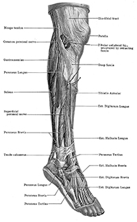
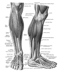
In other words, “accuracy” for Grant did not necessarily equal photographic realism; rather, it was a matter of truth to the holistic anatomical relationships being depicted. Many of Joy’s diagrammatic drawings were based on concepts that Grant used in his blackboard drawings. Figure 3 demonstrates Joy’s use of Grant’s approach to illustrate the three parts of the axillary artery and its branches. Joy recalls that she would constantly rework each drawing until the number of lines used to convey an idea was absolutely minimal (Joy, pers. comm., 2001). Grant’s Atlas of Anatomy was first published in 1943, and contained a wider variety of illustrative techniques than his Method of Anatomy. Grant’s objective in developing the Atlas was to produce a dissecting-room companion based on the specimens that students studied from in the Anatomy Museum at U of T. The date of publication is significant: before WWII, German anatomical atlases such as those by Carl Toldt, Johannes Sobotta, and Werner Spalteholz anchored medical curricula in North America as well as in Europe (Rosse 1999). The supply of German anatomical atlases on this continent was compromised by the war; the time was ripe for an indigenous publication. Grant’s Atlas both supplied this need and initiated a new approach to anatomical visualization based on realistic depictions of individual specimens (Figure 4), rather than on the idealized anatomy characteristic of the German atlases (Figure 5). The illustrations were painstakingly drawn from Grant’s dissections, and checked against other specimens to ensure accuracy (Joy, pers. comm., 2001). The following excerpt from the preface to the first edition of the Atlas describes the process that Grant directed his artists to use:
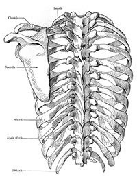
Thus, in the case of the Atlas, “accuracy” did mean something approaching photographic truth. Joy was responsible for most of the line drawings in the Atlas of Anatomy. Her pen and ink illustration of the rib cage (Figure 6) illuminates her mastery of this medium, through her gestural strokes and varying line weights. Although both Joy and Dorothy Foster Chubb contributed to the tonal illustrations, most of the tonal work was done by Chubb (Grant 1947). Grant’s views on medical art profoundly shaped the way that Joy approached her own work and, later, the teaching of medical illustration, as she acknowledges in an endnote to an editorial written for the Annals of Surgery in 1962. In this short article, she outlines a three-part taxonomy of illustration, consisting of diagrams, schemes, and lifelike approximations:
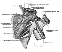
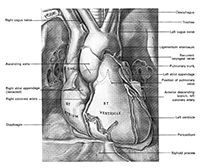
In the description of the diagram, we recognize the principle guiding some of Joy’s illustrations for Grant’s Method. In that of lifelike approximation, we see the principle guiding the illustrations for Grant’s Atlas. Grant’s books are still some of the most widely used texts in anatomy, and Joy’s contribution to these important works is immense. Her work was highly respected by Grant, who in the preface to the sixth edition of A Method of Anatomy pays tribute to “the cheerful, painstaking and talented services of Miss Nancy Grahame Joy” (Grant 1958). Most of Joy’s original artwork from Grant’s publications now resides in the archives of the Biomedical Communications program at U of T. Other freelance work, and entry into academia Joy worked briefly with Dr. C. G. Smith, a colleague of Grant’s who contributed a chapter to Morris’ Human Anatomy, 11th edition (1953). Figure 7 shows the dynamic approach that Joy took to illustrating the scapular muscles for this publication, and demonstrates her mastery of tonal watercolor illustration, which she perfected after being inspired by the work of Tom Jones. From 1952 to 1954, Joy freelanced in England and Scotland, including illustrations for Clinical Anatomy of the Heart (1978), which she considers to be particularly noteworthy (Figure 8) (Joy, pers. comm., 2001). In 1954, Joy returned to Toronto to continue her work with Grant. Two years later, in 1956, she accepted a position as a medical artist at the University of Manitoba in Winnipeg. (J.C.B. Grant had been Chair of Anatomy at the University of Winnipeg from 1919 to 1930; perhaps Joy’s decision to relocate there also reflects Grant’s influence.) Joy contributed to many publications during her time in Winnipeg, and was promoted there to her first academic position as Assistant Professor in the Department of Surgery. She considers this appointment the most important step in furthering her career in academia, since her status changed from non-academic to academic (Wilson-Pauwels 1993). Chair of Art as Applied to Medicine (AAM) In 1962, Maria Wishart (1893-1983) retired as director of the Art as Applied to Medicine (AAM) program at U of T, and Joy succeeded her as head of the department at the rank of associate professor. Wishart’s and Joy’s paths had long overlapped. The Wishart family and the Primroses, Joy’s maternal grandparents, were fellow parishioners at St. Andrew’s Presbyterian Church in downtown Toronto; Joy recalls visiting Maria Wishart as a teenager to seek advice about pursuing a career in medical art (Joy, interview, 1973, U of T Archives). It was Joy’s grandfather Alexander Primrose, during his tenure as Dean of the Faculty of Medicine at the University of Toronto, who had approved Maria Wishart’s appointment as the university’s first professionally trained medical artist in 1925 (Gilbert et al., 1995). Wishart had studied medical art under Max Brödel at Johns Hopkins Medical School from 1922 to 1925. In Toronto, she worked with other Hopkins-trained artists as her assistants and apprentices, including Dorothy Foster Chubb and Eila Hopper Ross. The three-year diploma course that she inaugurated in 1945 combined elements of apprenticeship and academic study; 19 students graduated from the program under Wishart’s leadership (U of T Archives). When Joy took over the program, the number of graduates each year was dwindling: only two Art as Applied to Medicine (AAM) diplomas were awarded between 1959 and 1964 (Gilbert et al., 1995). Joy knew that the AAM diploma program must be elevated to degree-granting status in order to survive (Joy, pers. comm. 2001). In 1965, she sat on an advisory committee to review the program; it was determined that “a diploma was not an adequate incentive or reward for the long years of training,” as she wrote in a retrospective summary to the Faculty of Medicine’s associate dean in 1973 (U of T Archives). Accordingly, in 1967, the AAM program was elevated to a three-year undergraduate Bachelor of Science in Art as Applied to Medicine (BScAAM) degree. Enrolment increased dramatically: by the time Joy retired in 1986, 76 students had received their BScAAM (Gilbert et al., 1995). Joy fought continually to maintain the department in the face of the university’s mounting financial pressures. She worked to further the program by promoting to other faculties the value of visual communication as a scholarly endeavor. In a speech to the Institute of Biomedical Engineering, for example, she declared:
She had struck a similar note in a departmental position paper in 1980, when she called for “AAM’s integration into all levels of the university” by “seeking formal academic recognition that to transmit information pictorially at a graduate level can be as significant an intellectual achievement as to do so in writing” (U of T Archives). Then, as now, medical artists had to fight for recognition of their discipline-specific knowledge base. As an educator, Joy kept ahead of the times in terms of technology. In 1971, for example, she completed a certificate extension course in Television Studio Production at the Ryerson Polytechnical Institute in Toronto and, that same year, helped to organize a Health Science Film Festival Workshop at U of T to promote awareness of the value of medical teaching films (U of T Archives). In 1982, she introduced a new course in the design of computer-aided learning for medical education, and obtained Telidon technology for the AAM program. The Telidon computer had 64K of memory, and could produce basic graphic shapes in eight colors. With this cutting-edge technology the students were able to create an interactive program for medical students to learn the pathways of spinal nerves (Lax, pers. comm., 2001). Advocacy Joy produced work as a freelance artist for most of her career, including her work for Grant. Neither Joy nor Chubb own any rights to their work in Grant’s publications, and Joy’s experience with various publishers led her to become a strong advocate for re-use fees (Joy, pers. comm., 2001). She introduced a course on business management to her students in AAM, and tried to educate other artists and authors through a 1964 article in Medical and Biological Illustration where she states that “copyright notice is especially necessary in the medical literature which is frequently abused by blatant plagiarism” (Joy 1964: 92). In a series of letters exchanged with a representative of U of T’s Scientific Development Committee in 1962, she had sought policy to define and protect the intellectual property rights of service and teaching staff and students in Art as Applied to Medicine (U of T Archives). Professional associations Joy joined the Association of Medical Illustrators (AMI) as an elected member in 1950. She served on the AMI’s Bibliography Committee as a member and later as chair, was an Associate Editor for the Journal of the Association of Medical Illustrators (1957-1963), sat on the AMI’s Board of Governors (1960-1965), and participated in the AMI’s Council on Education (1964 onward). Joy was also a charter member of the Canadian Academy of Medical Illustrators (1965-1981) and was awarded an Honorary Fellowship from the Ontario College of Art in 1984 for her contribution to the arts (BMC Archives). The AMI honored both Joy and Dorothy Foster Chubb at their annual meeting in 1998 in Toronto, when the publishers of Grant’s Atlas presented the original artwork used in the book to the Department of Anatomy and the Division of Biomedical Communications in the Department of Surgery. Legacy Joy stepped down as Chair of Art as Applied to Medicine in 1985, and retired from U of T one year later. Challenging times for the program still lay ahead—challenges that have been met by program directors Linda Wilson-Pauwels (1986 to 2008) and Nicholas Woolridge (2008 to the present). Under the leadership of Wilson-Pauwels, the program was renamed Biomedical Communications in 1990, to reflect its expanding academic and technological scope, and was elevated to the graduate level in 1993, so that graduates now receive a Master of Science in Biomedical Communications (MScBMC). Nancy Joy’s work in promoting Art as Applied to Medicine and elevating it to degree-granting status in 1968 contributed to the program’s growth. To this day, Joy is admired as a strong, independent visionary and consummate medical artist by the faculty, students, and alumni of this distinguished program.
References Allen, Cheralea Waite. 1983. Professor Nancy Joy: Marrying art and medicine. Alumnus [newsletter of the Ontario College of Art Alumni Association], Spring. Biomedical Communications Archives. Nancy Joy file. University of Toronto at Mississauga. Canadian Who’s Who, 18th ed. 1983. Toronto: University of Toronto Press. Gilbert, S., M. Mackay, L. Wilson-Pauwels and N. Woolridge. 1995. Art as Applied to Medicine/Biomedical Communications: Fifty Years of Growth. Toronto: University of Toronto Faculty of Medicine. Grant, J. C .B. 1943. An Atlas of Anatomy, 1st ed. Baltimore: Williams & Wilkins. Grant, J. C .B. 1947. An Atlas of Anatomy, 2nd ed. Baltimore: Williams & Wilkins. Grant, J. C. B. 1956. An Atlas of Anatomy, 4th ed. Baltimore: Williams & Wilkins. Grant, J. C. B. 1958. A Method of Anatomy, 6th ed. Baltimore: Williams & Wilkins. Joy, N. 2001. Interview by Jennifer A. Polk. Tape recording. Toronto, Ontario, 25 March. Joy, N. 1962. Pictured fact and fancy in current medical literature. Annals of Surgery 156(3): 511-512. Joy, N. 1964. Medical illustrations and copyright. Medical and Biological Illustration 14(2): 89-95. Joy, N. 1974. Occupational Information Monograph: Medical Illustrator. Toronto: Guidance Centre, Faculty of Education, University of Toronto. Joy, N. 1984. Creating the visual image. Speech presented at seminar for staff and students of Institute of Biomedical Engineering, 2 February. Lax, L. 2001. E-mail correspondence with Jennifer A. Polk. Toronto, Ontario, 24 April. National Archives (United Kingdom). Service record of Ernst Grahame Joy. Catalogue reference AIR 76/267; file reference 119. Robinson, C.L.N. 1988. Further remembrances of that revered anatomist, Dr. J.C. Boileau Grant. The Canadian Journal of Surgery 31(3): 203-204. Rosse, Cornelius. 1999. Anatomy atlases. Clinical Anatomy 12: 293-299. Schaeffer, J. Parsons, ed. 1953. Morris’ Human Anatomy: A Complete Systematic Treatise, 11th ed. New York: Blakiston. University of Toronto Archives, Department of Art as Applied to Medicine fonds [collection] 1926-1984, Accession A1985-0010/001-004. University of Toronto, “Faculty Council Minutes” Faculty of Medicine, University of Toronto, Toronto, Ontario, September 17, 1975. Wilson-Pauwels, L. 1993. The development of academic programs in medical illustration in North America from 1911 to 1991. Doctoral thesis, University of Toronto
Jennifer Polk, BSc, MScBMC, graduated from Biomedical Communications at the University of Toronto in 2002. She is currently Medical Media Design Coordinator for www.AboutKidsHealth.ca, a patient education website at The Hospital for Sick Children in Toronto. jen.polk@utoronto.ca Shelley Wall, AOCAD, MScBMC, PhD, is a faculty member in the Biomedical Communications program, University of Toronto Mississauga, where she teaches bioscientific illustration and research methods. Medical illustration in the mid-twentieth century is one of her research interests. s.wall@utoronto.ca
|
|
Copyright
2010, The Journal of Biocommunication, All Rights Reserved |