|
|
|
Visualization of Forensic Demonstrative Evidence for the Canadian Courtroom: A Collaborative Initiative between Biomedical Communications and Forensic Anthropology |
| Leila Lax, B.A., B.Sc.AAM, M.Ed.
|
AbstractThe 2004 expansion of the Biomedical Communications program at the University of Toronto at Mississauga provided new opportunities for inter-faculty teaching and re-conceptualization of the graduate curriculum, as well as professional practice outcomes. Collaboration between faculty from Biomedical Communications and the Department of Anthropology resulted in innovations in non-linear media design for forensic demonstrative evidence. Visual media experiments reinforced faculty collaboration that led to the expansion of an existing Medical Legal Visualization course, in the Master of Science in Biomedical Communications program. BackgroundThe 2004 expansion of the Biomedical Communications (BMC) program at the University of Toronto at Mississauga (UTM) provided new opportunities for inter-faculty collaboration and the re-conceptualization of the graduate curriculum, as well as professional practice outcomes. BMC faculty collaborated with a forensic anthropologist in the Department of Anthropology at UTM, to develop visualizations for forensic demonstrative evidence in the context of Canadian criminal law. Previously, BMC faculty only focussed on teaching development of demonstrative evidence for medical malpractice and personal injury cases. In Canada, medical malpractice and personal injury cases are tried in the civil courts of law. Therefore, in this paper, the term “medical legal” is used to refer to both medical malpractice and personal injury demonstrative evidence. Criminal cases are tried in criminal courts of law, and the term “forensic” demonstrative evidence is used to make this distinction.This paper will review current practices and paradigms of medical legal (medical malpractice and personal injury) and forensic visual evidence as used in Canadian courtrooms and at pretrial proceedings, and provide recommendations for future development. It also will document how interdisciplinary collaboration, between a forensic anthropologist and BMC faculty members, created a foundation on which to expand an existing Medical Legal Visualization course to include forensic visual evidence, signified by the new course title, “Visualization of Demonstrative Evidence for the Courtroom.” IntroductionMedical legal and forensic visualizations for demonstrative evidence are intended to support expert verbal testimony with didactic visual explanations, to clarify complex medical and scientific issues, to enhance understanding and affect more just decision-making and better legal outcomes. Studies regarding the effectiveness of visual evidence indicate that:People remember what they hear better when visual displays accompany the spoken word; after 72 hours, people remember only 10 percent of what they are told but 20 percent of what they are shown. When visual displays are used in the courtroom, however, the retention factor is 65 percent (Weiss-McGrath Report, 1986). Most people in a courtroom, including judges, jurors, and attorneys, have not been educated in the health sciences and therefore find medical and scientific terminology difficult to understand. The average educational level of jurors in Canada is grade eleven (Goldstein, 2003). Visual evidence is targeted to this level. Visual evidence can improve clarity and help overcome barriers to understanding. The power of visual persuasion is often cited by lawyers. “Visual evidence helps win cases because it improves judge and jury retention, highlights the salient points of a party’s case, recreates critical events in the evidentiary chain, and tells the story quickly and clearly” (Goldstein, 2003). Medical legal and forensic visualizations used as demonstrative evidence in the Canadian courtrooms must be accurate schematic representations essentially devoid of bias and interpretation. Development of visualizations for demonstrative evidence is based on primary visual evidence, i.e. radiographic and photographic images, medical records, and expert reports. It is important to note that primary visual evidence in most personal injury and medical malpractice surgical cases differ significantly from primary visual evidence usually available in forensic cases. Primary visual evidence in personal injury and medical malpractice surgical cases is often provided as a series of radiological images taken over time, i.e. x-rays, cat scans, and MRIs. Primary visual evidence for the creation of forensic visualizations for demonstrative evidence is most often provided in the form of photographs. At the crime scene, police and a forensic anthropologist photograph the bodily remains and the surrounding environment. The forensic anthropologist uses scientific methods to analyze the skeletal remains and their context at the scene and in the lab (Burns, 1999; Galloway, 1999). The skeletal remains are examined, tested, and often further photographed or x-rayed. Upon conclusion of the analysis, and usually before the trial, the skeletal remains are returned to the family for cremation or burial. The forensic anthropological findings are documented in an expert report providing information on reconstruction of the scene, the condition of the remains, the identity of the victim, and opinion as to the manner and mode of death. The medical visualization specialist uses the forensic expert report and the primary visual evidence, i.e. photographs and radiological images, in combination with feedback from the expert witness and lawyer to create accurate and admissible illustrations and animations. The overall development process and professional practice paradigms for forensic demonstrative evidence are analogous to those for medical legal. Medical legal and forensic visualizations, i.e. illustrations and animations, for the courtroom are categorized as demonstrative evidence and are described in the Canadian legal literature as illustrative or substantive according to the theoretical basis of each (Goldstein, 2003; Sopinka, 1997). According to the Illustrative Theory, visual evidence can be used only “to support, corroborate and explain the evidence of witnesses; to supply relevant detail (minutiae) in the appearance of objects described in oral testimony; to reveal steps taken by witnesses to arrive at their opinions; and, to affect the credibility attached to a witness’ testimony” (Goldstein, 2003). Illustrative demonstrative evidence is considered “pictorial testimony” and is inextricably linked to the testimony of an eyewitness. In trials for which there is no eyewitness to verify the visual evidence the Canadian courts adopt the Silent Witness Theory. Under this theory the courts consider visual demonstrative evidence as substantive evidence that can be authenticated by an expert witness, as a true and accurate depiction and thereby admitted to the court. Admissibility criteria for demonstrative evidence visualizations are similar in Canadian civil and criminal courts. These criteria were established in the case of R. v. Mohan (Supreme Court of Canada) and are similar to the American Daubert principles. Canadian criteria for admission of visual evidence are: When considering admissibility of demonstrative evidence in both Canadian civil and criminal courts, it is imperative that the probative value exceed the prejudicial effect. This means that the visualizations must not be inflammatory, be overly gruesome, or overemphasize any particular point or aspect portrayed. Accuracy and clarity of visual information is paramount for probative value. Once accepted into evidence, visualizations are demonstrated, or more often intrinsically woven into the context of expert oral testimony. Current Practice in Medical Legal Demonstrative EvidenceThe power of visualizations used as demonstrative evidence is well acknowledged in the legal community (Gelinas and Olah, 1997; Oatley, 1997). Medical legal visualizations, media and formats for presentation of demonstrative evidence used in preliminary hearings, mediation meetings, and in the courtroom, have evolved over the last 20 years in Canada, particularly in the Toronto region. Dorothy Irwin and Leila Lax are credited with initial development of the profession in this region in the mid 1980s. They were followed by Stephen Mader in the 1990s, whose studio currently employs many BMC graduates, and others who freelance in the field. Lax acknowledges the influence the Association of Medical Illustrators and her American counterparts in her initiation of the field. For many years large scale hand-painted, mechanically labeled, medical legal illustrations were the standard format (Irwin, 1997; Lax, 1997; Mader, 1996). In the mid 1990s, the introduction of computers and visual software led to the use of digitally created medical legal illustrations that were printed in large scale for the courtroom. In 1997, medical animation on VHS videotape for courtroom presentation on large screen televisions was introduced (Lax, 1997). Digital Static and Linear MediaOver the last decade in Canada, computer presentations have become increasingly popular for use in pretrial conferences, preliminary hearings, and the courtroom. Linear digital 2D and 3D animations and visualizations in PowerPoint® presentations are displayed using a laptop computer and projection equipment. Large scale digital prints and small scale digital handouts from animations and PowerPoint presentations are now commonplace and provide convenient ways to review issues for participants at a preliminary hearing or jurors in deliberation. The traditional format of medical legal illustrations, produced as large scale panels, remains ever popular; it will probably continue to be popular for cases with modest budgets and simple visual communication needs. The digital revolution has enabled many advances in media design for visual evidence. However, it is important to recognize the specific limitations of each format. Static digital illustrations and linear digital animations have significant limitations for presentation of demonstrative evidence in expert testimony. Static digital illustrations provide visual representation of only one perspective, which limits explanations of complex medical situations. This problem is often solved through the use of 2D and 3D animation, which has its own inherent limitations. Animations on videotape or on computer are presented in a linear sequence, from start to finish. Review of specific sections is difficult in these formats. PowerPoint presentations inherently have similar linear sequencing limitations. Expert oral testimony, and particularly testimony on cross-examination, does not usually follow a linear format or sequential storyline. In complex cases that require much visual evidence, linear formats are of limited use. As visual evidence needs and expectations become more sophisticated, it becomes apparent that a more effective format of visual demonstrative evidence is required to overcome these issues.
Nonlinear, Instant, Random Access MediaNew media and new methods of design were necessary to support nonlinear, instant, random access of visualizations to support effective didactic visual and verbal testimony for expert presentation and cross-examination. This design challenge was taken on in the BMC master’s Medical Legal Visualization course. Lax and her 2002 and 2003 students explored these issues, using Macromedia Flash® MX, with Jodie Jenkinson. Design innovations and outcomes for demonstrative evidence presentation, such as nonlinear, user-controlled interface design and radiographic-toimage “slider” visualization tool are detailed elsewhere (Lax et al., 2004).The advantages of nonlinear media design for demonstrative evidence requires further explanation. First, provision of multiple perspectives in a multimedia presentation, linking concepts from medical reports, radiographs with medical illustrations, and/or animations can help a lay audience, i.e. the jury, to connect concepts for deeper understanding. Second, unlike linear media, nonlinear interface design allows the expert witness to navigate, control, and adjust the visual presentation as needed, providing flexibility in presentation. As indicated above, this flexibility enables the expert witness to coordinate visual and verbal testimony. Synchronous testimony helps ensure simultaneous viewing and listening of evidence by the jury, the expert witness, and others in the courtroom. Dual coding theory indicates that simultaneous presentation of visual and verbal information results in cognitive processing for deeper understanding, enhanced memorability and recollection (Paivio, 1971; Clark and Paivio, 1991), all of which are important in jury deliberations and trial outcomes. Current Practice in Forensic Anthropology Demonstrative EvidenceVisualizations of demonstrative evidence are not commonly used by Forensic Anthropologists in Canadian criminal courts. However, photographs are often provided in booklet form. Forensic anthropologists usually title and label photographs of the skeletal remains. The photographs are organized in pages, and the booklets are submitted as demonstrative evidence. The photographs in the booklets are used to assist forensic anthropologists in describing findings at preliminary hearing or at trial. In the courtroom an individual booklet is provided for the judge, defense, and Crown attorneys, and a few copies typically are shared by jurors. The photographic booklets may provide anatomical context and visualizations to assist in complex scientific explanations of issues, such as manner and mode of death or weapon identification, but typically will focus on photographs of remains taken during analysis. The photographic booklet format is limited in media and design format for demonstrative presentation. The need for advancement of the current state of visual evidence as used by forensic experts in criminal cases is clear. Visualization Experiments for Future Practice in Demonstrative EvidenceThe defined limitations and need for advancement provided a common goal
for interdisciplinary collaboration between a Forensic Anthropologist
and BMC faculty. A series of visualization experiments were conducted
by Meaghan Brierley and Tracy Rogers, Ph.D., in collaboration with the
authors of this paper. The experiments examined the extrapolation of conceptual
constructs from the medical legal (Lax et al., 2004) to the forensic demonstrative
evidence context. Results from these visualization experiments were presented
at the Canadian Association of Physical Anthropology conference in London,
Ontario, Canada (Rogers and Brierley, 2004). Three visualization tools
were created to meet specific needs of forensic demonstrative evidence
and are described in detail below. They are: Slider VisualizationThe slider visualization tool aided in demonstrating the importance of frontal sinuses and suture patterns to the identification of skeletonized human remains. A similar principle was used to develop a visualization tool to promote shape recognition between photographs of human and nonhuman remains. In the first example, a slider is used to reveal the corresponding surface anatomy to radiographs of a human skull. By dragging the slider to the right, facial features gradually became more visible. The visual information of the first image, the radiograph, receded. The process of incremental transparency allows the expert witness complete control of the pace of the reveal. The lawyer or expert witness may repeat the action as many times as necessary to ensure visual comprehension by the jury. Once the viewer understands the relationship of the radiograph to the
face, a “Highlight Sinuses” button activates outlines of the
frontal sinuses (Figure 1). Comparison between frontal sinus patterns
can then be easily demonstrated for confirmation of personal identification
(Christensen, 2004). The outline of the sinuses can be turned on or off
at any time, and at any stage of image transparency. 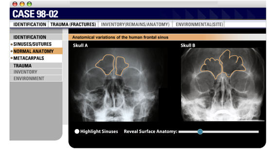
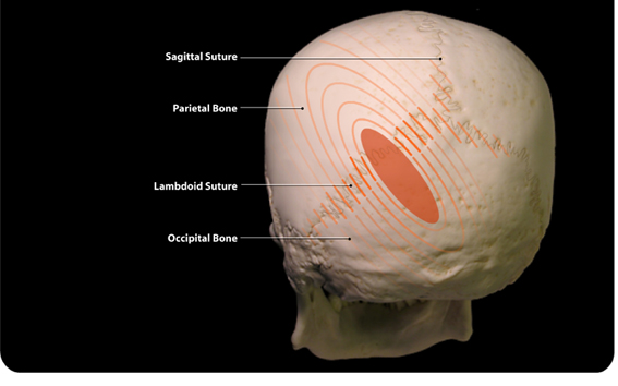
Figure 1. The slider is paused for comparison of frontal sinus patterns. Visual correlation of surface and deep anatomy with radiographic images also is possible. A variation of the visualization slider was used to merge two radiographs,
one on top of the other, to demonstrate that the cranial suture patterns
were the same. A "Highlight Sutures" button allows the user to emphasize
the pattern of the sagittal and the lambdoid sutures (Figure 2). Visual
comparison of cranial sutures is another important method used in the
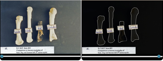
positive identification of human remains (Rogers and Allard, 2004). 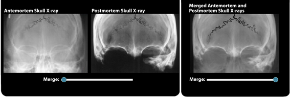 Figure 2. Cranial suture comparison between radiographs aided by the slider visualization tool. In Figure 3, the goal of the forensic anthropologist was
to prove that item #83 was a remnant of a human third metacarpal, as opposed
to a bone remnant of another species. A gradual reveal of the outlines
of the featured specimens, highlights the importance of bone morphology
and facilitates shape recognition.
Figure 3. Gradual reveal of outlines
to facilitate shape recognition.
Animated VisualizationsFigure 4 demonstrates the pattern of blunt force trauma to the skull using 2D animation overlays. The animated overlay indicates a blow to the skull across the lambdoid suture with a large blunt object. The dissipation of force from the blow is indicated along the suture and over the occipital and parietal bones. The forensic anthropologist would use this type of visualization to explain that a large instrument was involved since damage is apparent on both sides of the lambdoid suture, causing injury to both the occipital and the parietal bones. As indicated in this experiment, animated visualizations can be used to help explain type of trauma, area of impact, and size of the weapon.
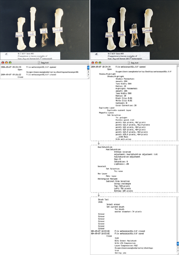
History LogsAdobe Photoshop CS has the ability to record a history log of interventions made to digital images. This function must be enabled by the user in the Photoshop CS preferences. Figure 5 shows two photographs with varying degrees of intervention, and the corresponding text that was recorded with each intervention. The first file (a) was opened and closed; the text recorded and represented here is very short. The second file (b) was opened, its shadows, highlights, hue and saturation adjusted, and a white line was painted around to edge of each of the specimens. This log is visibly longer. These log files can be printed and submitted with the visual demonstrative evidence, as supporting documentation. It is important to note that all photographic enhancements should comply with the American Federal Bureau of Investigation guidelines. These guidelines, found at http://www.fbi.gov/hq/lab/fsc/backissu/jan2003/swgitdigital.htm (FBI, 2003), and other digital evidence recommendations and guidelines for admissibility can be accessed in the online journal, Forensic Science Communications. We encourage use of the American guidelines by Canadians, since we are not aware of any in current publication pertaining to Canadian digital evidence.
Figure 5a. Log indicates no changes
were made to this image.
Figure 5b. Log of this photograph reveals that its shadows and highlights were adjusted, areas were selected to make hue and saturation changes, and finally, a brush tool and eraser tool were used to paint a white line around the specimens. In summary, results from the forensic visualization experiments indicate that:
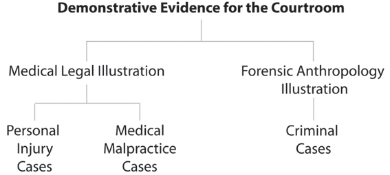
Discussion and Implications of the Interdisciplinary CollaborationIt became apparent that the advancement of visualization ideas in one area, in the medical legal or in the forensic context, can provide stimuli for extrapolation, to mutually inform and advance both fields of practice. Various constructs developed in the Medical Legal Visualization course were successfully extrapolated to the forensic demonstrative evidence context. These include the visualization slider and nonlinear, media formats for presentation (Lax et al., 2004). The forensic visualization experiments generated new ideas. Interactive shape recognition and animation overlays were shown to be effective ways to convey visual information. Digital history logs were shown to provide necessary and detailed records of change. The expansion of the BMC program to the UTM campus, Institute of Communication and Culture in 2004, by Director Linda Wilson-Pauwels, Ed.D., opened new avenues for collaboration between BMC faculty and UTM colleagues. Success of initial forensic visualization collaboration, translation of previous course constructs, and novel visualization experiment outcomes prompted reconceptualization of the existing Medical Legal Visualization graduate course in BMC. BMC faculty collaborated with Tracy Rogers, Ph.D., forensic anthropologist in the Department of Anthropology at UTM, to develop a forensic visualization stream to complement the medical legal visualization stream. Consequently the name of the BMC master’s course was changed to “Visualization of Demonstrative Evidence for the Courtroom.” Focus on personal injury and medical malpractice cases was expanded to include criminal cases (Figure 6). The course was implemented in the fall of academic year 2005. A complementary undergraduate course, titled Forensic Visualization for Demonstrative Evidence, is currently in development by the collaborators, and will be jointly offered through the UTM, Institute of Communication and Culture, new Health and Science Communication specialization and the UTM Department of Anthropology in January 2006.
ConclusionRecent collaboration between BMC and forensic anthropology faculty indicates that visualizations for demonstrative evidence developed for medical malpractice and personal injury cases can be successfully translated to the forensic visualization context, and, therefore, integrated in various academic paradigms. We anticipate that there will be many fruitful teaching, learning, and research opportunities, as well as professional practice outcomes, which will arise from continued collaboration. As demonstrated in this initiative, interdisciplinary emergent ideas can expand the knowledge bases of both disciplines, forensic anthropology and BMC. ReferencesBurns, K.R. 1999. Forensic anthropology training manual. Upper Saddle River, NJ: Prentice-Hall, Inc. Christensen, A. M. 2004. The Impact of Daubert: Implications for Testimony and Research in Forensic Anthropology (and the Use of Frontal Sinuses in Personal Identification), Journal of Forensic Science 49(3):1-4. Clark, J. M. & Paivio, A. 1991. Dual coding theory and education. Educational Psychology Review 3(3), 149-170. Federal Bureau of Investigation. Forensic Science Communications, Standards and Guidelines. accessed 10.27.04 http://www.fbi.gov/hq/lab/fsc/backissu/jan2003/swgitdigital.htm Galloway, A. 1999. Broken Bones: Anthropological Analysis of Blunt Force Trauma, Springfield, IL: Charles C. Thomas Publisher Ltd. Gelinas R. and J. A. Olah. 1997. The Power of Demonstrative Evidence. In Technology and the Trial Lawyer, Conference Proceedings of the Canadian Bar Association, Continuing Legal Education: Toronto, ON. Goldstein, E. 2003. Visual Evidence: Practitioner’s Manual, Scarborough, ON: Carswsell Thomson Professional Publishing. Irwin, D. 1997. Maximizing the Use of the Medical Illustrator, Part I. In Demonstrative Evidence for the Courtroom, Conference Proceedings of the Ontario Trial Lawyers Association, Toronto, ON. Lax, L. 1997. Maximizing the Use of the Medical Illustrator, Part II. In Demonstrative Evidence for the Courtroom, Conference Proceedings of the Ontario Trial Lawyers Association, Toronto, ON. Lax, L., Taylor, I., Wilson-Pauwels, L. and Scardamalia, M. 2004. Dynamic Curriculum Design in Biomedical Communications: Integrating a Knowledge Building Approach and a Knowledge Forum® Learning Environment in a Medical Legal Visualization Course. The Journal of Biocommunication 30:1. Mader, S. 1996. The Preparation of Customized Medical Illustration Exhibits for use in the Courtroom. In Visual Evidence: Practitioner’s Manual, Goldstein, Elliott, Ed., Scarborough, ON: Carswell Thomson Professional Publishing, 16:18-44. Oatley, R. G. 1997. Computerization of Demonstrative Evidence: Changing the Face of Advocacy. In Demonstrative Evidence for the Courtroom, Conference Proceedings of the Ontario Trial Lawyers Association, Toronto, ON. Paivio, A. 1971. Imagery and Verbal Processes. New York: Holt, Rinehart & Winston. Rogers, T. L. and Brierley, M. 2004. Forensic Visualization for Expert Testimony. Presentation at the Annual Conference of the Canadian Association for Physical Anthropology, London, Ontario, Oct. 30, 2004. Rogers, T. L. and Allard, T. T. 2004. Expert Testimony and Positive Identification of Human Remains Through Cranial Suture Patterns. Journal of Forensic Science 49(2)1-5. Sopinka, J. 1997. It Looks Great, But Can You Use It? Admissibility Issues with Respect to Computer-Generated Evidence. In Technology and the Trial Lawyer, Conference Proceedings of the Canadian Bar Association, Continuing Legal Education: Toronto, ON. Weiss-McGrath Report. 1986. Using Graphics in Court: An Art in Itself. 22 Trial (ALTA Magazine)1(77). Acknowledgements:Jodie Jenkinson, B.A., M.Sc.BMC, Assistant Professor in Biomedical Communications, Institute of Medical Sciences at the University of Toronto, is acknowledged for the original programming of the visualization slider. The original slider was developed for Wiley, M., Stewart, P., Liebgott, B., Jenkinson J., Cohen, H. 2002. ANATOMIA: Functional human anatomy electronic resource, http://www.bmc.med.utoronto.ca/anatomia. The slider used for this series of visualizations was inspired by the object-oriented programming of freelance consultant Grant Skinner, Edmonton, Alberta, Canada. http://www.gskinner.com Authors:Leila Lax (B.A., B.Sc.AAM, M.Ed.) is an Assistant Professor in Biomedical Communications, Institute of Communication and Culture, University of Toronto at Mississauga, and an Associate Member of the Institute of Medical Science, Faculty of Medicine, University of Toronto. She is a senior research scientist with the Institute for Knowledge Innovation and Technology and a member of the Wilson Centre for Research in Education. Lax has received numerous teaching awards. Her research interests include the design and evaluation of knowledge building/knowledge translation in Internet-based collaborative learning environments for health professional education.Email: l.lax@utoronto.ca Tracy Rogers (B.A., M.A., Ph.D.) is a consulting Forensic Anthropologist and an Assistant Professor in the Department of Anthropology at the University of Toronto at Mississauga. Rogers has assisted the police and coroner’s office in more than 150 forensic cases, including the investigation of the property of alleged serial killer Robert Pickton in Coquitlam, British Columbia. Rogers is actively involved in developing forensic anthropology in Canada and has published on topics related to personal identification (sex, age, ancestry, identification theory), crime scene investigation, and ethics. Meaghan Brierley (B.F.A., PGDip. Medical Photography, MScBMC) is a Research Associate with the IVIS (Interpretive Visualization) Group and an instructional media designer and developer with the Bell University Lab in Health Communication, Faculty of Medicine, U of T. Brierley is a research, teaching assistant and new media consultant in the graduate course in Demonstrative Evidence at U of T. Linda Wilson-Pauwels (AOCA, B.Sc.AAM, M.Ed., Ed.D.) is Director of Biomedical Communications, Institute of Communication and Culture at UTM and Director of the Master of Science in Biomedical Communications program, Institute of Medical Science, Faculty of Medicine, U of T. Wilson-Pauwels is a tenured professor in BMC and is cross-appointed to the Department of Surgery. She is a visual communicator with advanced degrees in higher education. Her interest is in information transfer to target audiences, development of visual organizational tools, and visual cues and their role in directing attention. Margot Mackay (ANSCA, B.Sc.AAM) is a tenured Professor
in Biomedical Communications, Institute of Communication and Culture,
UTM and an Associate Member of the Institute of Medical Science, Faculty
of Medicine, U of T. Mackay holds a cross appointment in the Department
of Surgery and teaches Surgical Illustration, a unique course in which
process and story telling are portrayed via sequential visualization.
|
Copyright 2004, The Journal of Biocommunication, All Rights Reserved