|
| Calendar & News | Columns | Features | Gallery | Showcase | Archives |
|
|||||||||||||||
|
Ophthalmic Imaging - an Overview and Current State of the Art, Part II |
| Timothy J. Bennett, CRA, FOPS In the 1960’s, a number of technological advances in photography helped to usher in a new era of diagnostic testing in ophthalmology, and with it a new profession was born. Ophthalmic photography is a diagnostic discipline that utilizes a number of photographic techniques to help document and diagnose various diseases of the eye. The photographers that formed the vanguard of this new profession found a shared need to exchange information, collaborate on new techniques, and set standards of practice in the profession. They formed the Ophthalmic Photographers’ Society, Inc (OPS) to fill these needs. Through the collaborative efforts of OPS members, the profession has grown and flourished. Today, OPS members continue to promote and elevate the profession through publication, education, and certification. Changing health care delivery models and new technological developments have altered the shape of the profession in recent years. Many photographers have expanded their job responsibilities and developed new skills in related fields. As new diagnostic imaging and treatment modalities are developed, the role of the ophthalmic imager will evolve and continue to play an important role in the preservation of sight. Part two of this overview extends beyond photographic technique and explores the evolution of ophthalmic photography as a profession.
|
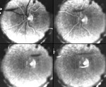
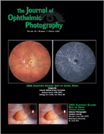
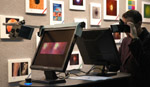
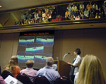
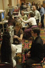
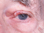
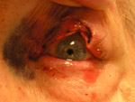
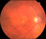
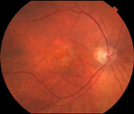
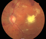
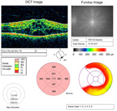
Ophthalmic Photography as a Profession Although there are published accounts of ophthalmic photographic techniques going back to the late 1800s, ophthalmic photography as a profession didn’t evolve until the 1960’s. A number of historical events coincided to provide the foundation for practical retinal photography. Kodak Tri-X film was introduced in 1954. The following year, the Zeiss-Littman modern fundus camera was introduced. It was the first commercially produced fundus camera to incorporate electronic flash illumination (Van Cader 1978). In 1959, two medical students, Harold Novotny and David Alvis started a research project to develop a photographic technique to estimate blood oxygen concentrations in the retinal vasculature as a visible segment of the cerebral circulation. In doing so, they worked out the basic techniques of fluorescein angiography and performed the first successful angiogram of a human retina (Figure 1). They reported their results in a landmark paper in 1961 (Novotny and Alvis 1961). Following their report, popularity of the technique exploded, as several investigators studied various clinical applications of angiography, quickly establishing the intrinsic value of the technique (David and Justice 1994). Up until this point, most ophthalmic photography had been performed by physicians, researchers, assistants, and in some cases, medical photographers. The sudden enthusiasm for fluorescein angiography created an immediate need for skilled full-time retinal angiographers, providing the foundation for the profession as we know it today. The first generation of “ophthalmic photographers” to enter the field were mostly medical photographers who already had some experience in fundus photography and were able to make a quick transition to this new subspecialty (Wong 1979). These early practitioners worked side-by-side with ophthalmologists and retinal specialists in exploring the diagnostic uses of fluorescein angiography and learning together as they unraveled the complexities of interpreting the fascinating images they were capturing. This close clinical collaboration with physicians quickly elevated the profession. Many ophthalmic photographers contributed significantly to the ophthalmic literature of the time and were held in high regard as professional colleagues in ophthalmology. This spirit of scholarly collaboration between photographer and physician continues today in many academic practice settings. Over time, there were gradual shifts in the professional experience of individuals entering the field. A second generation of fluorescein angiographers came from a cross-section of commercial, industrial, and scientific photography backgrounds, adapting their existing photographic skills to ophthalmic subjects. This was followed by another group of personnel that had experience in ophthalmology as ophthalmic technicians, but with no technical photographic training. The diversity of professional backgrounds of individuals entering the field created a universal need for education and sharing of information. With no real formal training available in ophthalmic photography, practitioners had to learn many necessary skills on the job. Although at least one current biomedical photography degree program offers elective courses in ophthalmic photography, the vast majority of practitioners in the field have no formal training in ophthalmic photography. In the absence of formal education in ophthalmic photography, it may seem incongruous that the profession provides a high level of academic pursuit and achievement for many members. A number of ophthalmic photographers contribute to the profession of ophthalmology as authors of ophthalmic textbooks and scientific articles, some participate as investigators or sub-investigators in clinical research, and many play an integral role as instructors in ophthalmology residency training programs. Through these contributions and academic achievements, some ophthalmic photographers have even received academic appointments as professors of ophthalmology. In the decade that followed the introduction of fluorescein angiography, some of the pioneers in the field occasionally crossed paths while attending ophthalmology meetings. In such a young profession, workers in the field found a great desire for interaction with fellow professionals to share experiences and techniques. In April of 1969, a group of photographers had an informal gathering during the Association for Research in Vision and Ophthalmology meeting in Sarasota, Florida to discuss forming a professional society (Justice 1978). They agreed to have their first formal meeting in Chicago during the American Academy of Ophthalmology meeting later that year. Attending the first meeting were ten ophthalmic photographers, who set organizational goals, selected interim officers and chose a name (Figure 2). The Ophthalmic Photographers' Society, Inc (OPS) was incorporated in July of 1970. The OPS is a non-profit organization that today counts over 1000 members from 27 countries. Membership is open to anyone with an interest in ophthalmic photography and is made up of photographers, technicians, physicians, scientists, vendors and students. The main objectives of the Society are to provide primary and continuing education in the field of ophthalmic photography, to set and maintain professional standards through certification, and to promote scientific advancement in imaging technology and techniques. Since its inception, the OPS has provided a central forum for the exchange of information through a number of programs and publications. The Society sponsors national and regional educational meetings, offers educational scholarships, publishes the peer-reviewed Journal of Ophthalmic Photography (Figure 3), as well as a member newsletter, maintains a website filled with important news and technical information, and offers certification in ophthalmic photography. All of these programs and member benefits are accomplished almost entirely through the efforts of dedicated volunteers from the OPS ranks, along with generous philanthropic support from our sustaining members. Despite the highly specialized nature of the profession, the OPS has found shared interests and collaborative opportunities with other organizations, such as NOCA, JCAHPO, ICOP, and others. The OPS has been a member of the National Organization for Competency Assurance (NOCA) since 1989. NOCA establishes standards for accreditation of certification programs and provides a forum for organizations interested in competency assurance and certification. The OPS is one of seventeen member organizations of the Joint Commission for Allied Health Personnel in Ophthalmology (JCAHPO), which provides education and certification for related allied health professions in eye care. The International Conference on Ophthalmic Photography (ICOP) is a joint venture of the Australian Institute of Medical and Biological Illustration (AIMBI), the Japanese Ophthalmic Photographers' Society (JOPS), the Ophthalmic Imaging Association (OIA) from the United Kingdom, the Ophthalmic Fotographers of the Netherlands (OFN) and the Ophthalmic Photographers' Society. Since 1986, this quadrennial scientific symposium has been held in different venues around the world. The OPS was the host organization for ICOP 2006 in San Francisco and has passed the torch to OFN, who will host ICOP 2010 in Amsterdam. The OPS also participates in the annual meetings of the American Academy of Ophthalmology and the American Society of Cataract and Refractive Surgeons, by sponsoring photographic competitions that showcase award-winning photographs in scientific exhibits at the annual meetings of these physician organizations. The stunning exhibits are a “must-see” highlight for both photographers and ophthalmologists attending these meetings (Figure 4). Education and Certification The diverse makeup of the OPS membership underscores the continuing need for education in the field. The OPS sponsors national, regional, and international education programs that provide comprehensive training opportunities for ophthalmic imagers. The OPS Annual Educational Program, held in conjunction with the Annual Meeting of the American Academy of Ophthalmology, is the premier educational opportunity in the field (Figure 5). The diverse curriculum includes offerings from entry-level techniques and patient care, to updates on the latest technology, advanced techniques, image interpretation, and electronic communication. In addition to the didactic and hands-on educational opportunities, a hallmark of all OPS programs are the social events that provide invaluable networking opportunities reminiscent of the initial gatherings of the founding members of the Society. The 38th Annual OPS Educational Program will be held in November, 2007 in New Orleans, LA. Education and certification go hand-in-hand in ophthalmic photography. OPS sponsored educational programs, when combined with certification, form a diverse curriculum for professional growth and education that extends well beyond "on-the-job training." The Certified Retinal Angiographer, (CRA), program was established by the OPS Board of Certification in 1979. To date, nearly 800 individuals have successfully achieved recognition as a CRA. This credential is recognized in the ophthalmic community as an objective measure of competence in fundus photography and fluorescein angiography, and is meant to assure employers and the public that an individual has demonstrated a high level of proficiency in the field. The CRA Program is accredited by the by the National Commission for Certifying Agencies (NCCA). NCCA standards are recognized in the professional certification and testing community as the benchmark for quality and fairness in competency assurance programs. Accreditation of the CRA program reinforces the value of the credential, and the respect associated with those who hold it. Continuing education is important in maintaining one’s skills and is a requirement for recertification as a CRA. To promote access to educational opportunities, the OPS has established a scholarship fund to help offset the costs for selected recipients to attend educational meetings in ophthalmic photography. Looking to the future, the OPS plans to expand continuing education programs through development of new distance-learning opportunities including online tutorials, podcasts, and “webinars.” In response to changes in job responsibilities identified by recent surveys, the OPS Board of Certification is currently developing a new certification program in optical coherence tomography (OCT), a relatively new technology that has revolutionized diagnostic imaging over the last several years. The program is scheduled for launch in the Fall of 2007. Current And Future Trends Digital Imaging In Ophthalmology Perhaps the most significant technological change in the profession since the early days of angiography has been the implementation of digital imaging. The transition from film-based imaging to digital technology in ophthalmology has been ongoing for over two decades. Commercial digital systems designed specifically for fluorescein angiography and retrofitted to existing fundus cameras began to appear on the market as early as 1983. Color digital imaging took longer to penetrate the market, and until recently film remained the medium of choice for color fundus and slit-lamp photography in most practice settings. Digital imaging offers some distinct advantages over traditional film-based imaging in ophthalmology. In addition to well-known advantages in capturing, enhancing, storing, displaying and distributing images electronically, having instant access to digital images can improve clinical efficiency and patient education. Digital analysis enables measurement of pathologic structures and digital overlays can be used to identify changes in lesion size in serial photographs. Perhaps the biggest advantage of digital imaging is that it can shorten the learning curve for novice photographers trying to master some of the more complex ophthalmic imaging techniques. Having instant feedback allows the photographer to quickly adjust alignment and camera settings to correct flaws in technique. Despite these advantages, the high initial cost of digital systems has prevented them from being employed universally, although they are now more common than film-based systems. A pair of surveys looking at tasks performed by retinal angiographers indicated that over 70% of respondents are utilizing digital technology for some or all of their fluorescein angiograms (Bennett, MacGregor, and Scheuneman 2004) (Benetz, Bennett, and Tomer 2005). The same surveys showed an increase in use of color digital imaging for fundus photography from 46% in 2002 to 73% in 2004. These numbers would surely be higher today. There are however, some disadvantages to digital imaging in ophthalmology. The digital revolution has surprisingly led to an overall decline in the quality and consistency of external photography. The ease of use of point-and-shoot digital cameras has made them popular for use by physicians or other non-photographers in clinical settings. These cameras are good tools for some uses, but the built-in flash offers no lighting flexibility for external eye photography. The autofocus zoom lenses lack distance markings or other methods to standardize magnification settings for repeatable results. Using the “macro” mode often disables the flash, leading to inaccurate color rendition from mixed ambient lighting (Figure 6). The macro mode may also set the zoom to its wide-angle setting, causing an elongation of perspective at close range. Users that understand the principles of focal length, depth of field, magnification and lighting can make reasonable use of the these cameras for external photography, but the digital SLR is clearly the best tool for the job. Unfortunately the trend is moving away from the routine use of SLRs. Another limitation of digital imaging is in its use for color fundus photography, which is probably the most commonly performed procedure in ophthalmic imaging. Most color sensors employed today do an excellent job in capturing a full spectrum of color for general pictorial use, but often have a difficult time accurately rendering subtle color differences in the red, orange, yellow range (Figure 7). These are the most important colors in fundus photography and color transparency films do an excellent job in rendering them accurately. Even the best color digital systems have yet to match the color accuracy of transparency films for color fundus imaging. There is also a lack of standardization in color, exposure, and image-processing settings, and the resulting images often appear oversaturated and oversharpened. Education and strong standards are needed to improve utilization of digital color imaging tools. Routine use of color management calibration tools will also improve the situation. Photographic reading centers that grade images for clinical research rely on standardized imaging protocols to obtain consistent photographs from multiple photographers and clinics. As the reading centers transition from film to digital submissions, they are starting to implement new standards to help resolve many color reproduction issues associated with digital fundus photos. The OPS will also play a key role in developing and disseminating new digital standards. Digital imaging has also had a huge impact in the presentation technology used for ophthalmic education. In just a few short years we have witnessed a dramatic change in the way that lectures are presented. The days of delivering ophthalmic lectures with side-by-side 35mm slide projectors have rapidly disappeared. Most major educational meetings no longer support 35mm projection, and now require all speakers to present electronically using LCD projectors and computer presentation software such as Microsoft PowerPoint™. Digital Imaging Integration As the move to an all-digital environment continues, ophthalmic images will increasingly need to be integrated into electronic medical records and picture archiving and communication systems (PACS). Commercially available digital angiography systems typically support the Digital Imaging and Communications in Medicine (DICOM) Standards, a set of comprehensive standards for handling, storing and transmitting diagnostic images. Conformance to these standards greatly simplifies the integration of imaging devices from various manufacturers with PACS and other hospital information systems (Bidgood and Horii 1992) (Kabachinski 2005). Ophthalmic imaging originally was included under the photographic visible light supplement to the DICOM standard, but in 2004 a new supplement introducing specific Ophthalmic Photography image classes was adopted. Although DICOM capability has been available for years, ophthalmology has been slow to adopt use of the standards. Unlike radiology services that are usually concentrated in hospitals or medical centers, ophthalmic imaging is performed in a wide range of practice settings. Large medical centers have the resources and infrastructure to make practical use of DICOM standards as a single point of integration for all diagnostic imaging modalities, while smaller ophthalmology practices may not. Diabetic Retinopathy Screening A recent trend in ophthalmic imaging is the use of digital screening for diabetic retinopathy. Diabetes is a significant worldwide health problem affecting an estimated 18 million people in the United States alone. One of the most devastating complications of diabetes is blindness from diabetic retinopathy. Early detection and treatment can significantly reduce the incidence of blindness from retinopathy; however, only about half the known patients with diabetes receive recommended annual eye examinations. The reasons for the low rate of patient compliance are complex, and include poor access to ophthalmologists in underserved areas. Digital fundus photography is an effective method of retinopathy screening that is capable of detecting macular edema and proliferative diabetic retinopathy, the most common causes of vision loss in diabetics (Figure 8). Non-mydriatic digital fundus cameras are designed with an infrared focusing system that promotes physiologic dilation in a darkened room, making them simple to operate. They can be placed at remote primary care sites and operated by available clinical personnel such as nurses and medical assistants. Images are directed to a centralized reading center for image grading and treatment recommendations (Merin, Guentri, Recchia 2004). Although professional ophthalmic photographers generally don’t perform imaging in digital screening programs, they often oversee network design, training and sometimes serve as primary image graders in screening programs. Digital retinopathy screening programs have been implemented in many locations around the world and are most successful in countries with centralized health care, such as the government sponsored screening program in the United Kingdom. Cross Training As diagnostic imaging has become ubiquitous in most ophthalmic practice settings, the roles and backgrounds of those performing photography has slowly shifted. More and more ophthalmic technicians and assistants have cross-trained to do some imaging procedures. Conversely, many ophthalmic photographers have gone on to obtain training in some of the skills required of ophthalmic technicians, further blurring the line between these two allied health professions. Cross-trained professionals who can perform patient screening work-ups as well as photography are highly valued and sought after by smaller ophthalmology offices. Because of the technical background required for many of today’s imaging procedures, some experienced ophthalmic photographers have taken a logical step and have assumed responsibility for computer network setup and maintenance for their practices, acting as a part-time IT specialist. One of the more controversial job responsibilities for many ophthalmic photographers is performing intravenous injections for fluorescein and ICG angiography. In some practice settings it is logistically advantageous for photographers to do injections themselves, as long as they have received documented training in venipuncture, IV administration of dyes, and universal precautions (Scott 1999). There can, however, be legal issues associated with unlicensed personnel performing fluorescein injections in some states (Ellis and Weber 1995). It is recommended that imaging personnel check their current state or local laws regarding the credentialing requirements of personnel performing intravenous injections (OPS 1999). Some cross training is probably inevitable in the field of ophthalmic imaging. Not all practice settings can support a full-time ophthalmic photographer, so cross-trained individuals have more employment options. There is however, still a place for dedicated ophthalmic photographers in academic medical centers and large retina or multi-specialty practices. Future of the Profession The future of ophthalmic imaging is promising but unclear. New technology brings new challenges as well as opportunities to advance health care and improve the quality of life for our patients. The field has witnessed technology- driven shifts in the utilization of diagnostic imaging procedures as new instruments have become available. For example, optical coherence tomography (OCT), a relatively new technology, is now one of the most commonly used diagnostic procedures in ophthalmology and has reduced the need for angiography for some conditions. Treatments in ophthalmology are also changing; and new or different diagnostic tools and strategies may be necessary to monitor these treatments. The movement is likely to continue as treatment of diabetic retinopathy and macular degeneration shift from traditional laser treatment to management with novel medications. New diagnostic instrument designs that offer automated image capture are currently being developed, with an emphasis on quantitative measurement and analysis rather than subjective image interpretation. There are some concerns about the future of the profession of ophthalmic photography as more automated instruments come to market. So far, imaging-based quantitative measurements have proven to be very dependent on image quality; and cutting-edge technology may require highly skilled operators to obtain consistent results (Figure 9). Another likely trend is the development of combination devices incorporating multiple imaging technologies in a single instrument. It remains to be seen whether these new technologies will require more, or less skill to operate, and how that will effect the profession. As the field continues to evolve beyond traditional photographic techniques to include more scanning-based imaging modalities, the perception of our job title has begun to evolve from photographer to imager. In response to this shift the OPS has adopted the identifying slogan, “Eye Imaging Experts”. Ophthalmic photography continues to be a rewarding career with many potential employment opportunities in the coming decades. Over the past several years, the OPS has conducted a number of member surveys designed to identify job tasks and gather demographic data for establishing continued job relevance of our certification programs in compliance with NCCA recommendations. These data have been instrumental in shaping the evolution of our education and certification programs. Each of these surveys was designed to provide a look at the tasks required in our profession from a particular vantage point in time. When viewed together, additional information becomes apparent when comparing demographic information that was captured with each survey. For example, the reported years of experience has steadily increased in direct proportion to the time elapsed between surveys. This indicates a stable number of very experienced professionals in the field. In 2004, one third of the respondents reported seven or more years of experience and nearly half reported more than 15 years of experience in the field. As a profession we don’t experience a high turnover rate that may affect other allied health professions. The data suggests that ophthalmic photography can be a rewarding, life-long career choice, but also suggests a cause for future concern. This experienced group is also getting older, and a large percentage will reach retirement age at a time that is likely to coincide with a widely predicted increase in demand for healthcare services. It is incumbent on the profession to look to the future to attract new recruits to the field, share the knowledge gained through experience, and provide both education and certification to the next generation of “Eye Imaging Experts.” The OPS remains a strong and vital professional organization despite many changes that have affected health care professions over the last decade. Our membership numbers and resources remain stable, our educational programs are exceptional in quality and diversity, the CRA program is accredited, and the Journal of Ophthalmic Photography has never looked better. These successes are a testament to the commitment of time, talent and energy of our members. Author's note: Reference/tutorials on Ophthalmic Photography techniques are available on the Ophthalmic Photographers' Society website at: http://www.opsweb.org/OpPhoto/OpPhoto.html. Searches can be performed on the Journal of Ophthalmic Photography at http://www.opsweb.org/Publicat/Journal/JourSrch.html. Citations are linked to PDF files containing the associated article. References Benetz BA, Bennett TJ, Tomer TL. What does today's ophthalmic photographer do? J Ophthalmic Photography 27:85-7, 2005. Bennett TJ, MacGregor G, Scheuneman JD. Digital Imaging and the certified retinal Angiographer Program. J Ophthalmic Photography 26:28-36, 2004. Bidgood WD Jr, Horii SC. Introduction to the ACR-NEMA DICOM standard. Radiographics 12:345-355, 1992. David NJ, Justice J. The early days of fluorescein angiography. J Ophthalmic Photography 16:83-86, 1994 Ellis JH, Weber P. Legal issues arise when unlicensed personnel administer IV fluorescein. Argus Nov/Dec:28-31, 1995. Friberg TR, Rehkopf PG, Warnicki JW, et al. Use of directly acquired digital fundus and fluorescein angiographic images in the diagnosis of retinal disease. Retina 7:246-251, 1987. Justice J. The ophthalmic photographers’ Society, a biographical sketch. J Ophthalmic Photography 1:5, 1978 Kabachinski J. DICOM: key concepts—part I. Biomed Instrumen Technol. 39:214-216, 2005. Kabachinski J. DICOM: key concepts—part II. Biomed Instrumen Technol. 39:292-294, 2005. Merin LM, Guentri K, Recchia CC. Digital detection of diabetic retinopathy — increasing access, reducing risk, improving outcomes. J Ophthalmic Photography 26:59-66. Novotny HR, Alvis DL. A method of photographing fluorescence in circulating blood of the human retina. Circulation 24:82-86, 1961. Ophthalmic Photographers’ Society Standards of Practice. J Ophthalmic Photography; 21:26, 1999. Scott JR. Dye Injection by Ophthalmic Photographers. J Ophthalmic Photography 21:7, 1999. Van Cader, TC. History of ophthalmic photography. J Ophthalmic Photography 1:7-9, 1994. Wong D. Editorial. J Ophthalmic Photography 2:3, 1979. Timothy J. Bennett, CRA, FOPS, is an ophthalmic photographer in the Penn State University Department of Ophthalmology at the Milton S. Hershey Medical Center. Mr. Bennett has over 25 years experience working in university-based academic medical centers. He is a nationally recognized author, lecturer and educator in the field of ophthalmic photography. Mr. Bennett holds current certification as a Certified Retinal Angiographer, is a Commissioner of the Joint Commission on Allied Health Personnel in Ophthalmology, and has been recognized for his contributions to the profession by being named a Fellow of the Ophthalmic Photographers Society. He has served on the OPS Board of Certification, the OPS Board of Directors, and is currently President of the Ophthalmic Photographers’ Society. Email: tjbennett@psu.edu |
Copyright
2007, The Journal of Biocommunication, All Rights Reserved
Table
of Contents for VOLUME 33, NUMBER 1