 |
||||
Viewpoint: Recapturing Science in Clinical Photography |
David Teplica
|
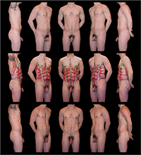
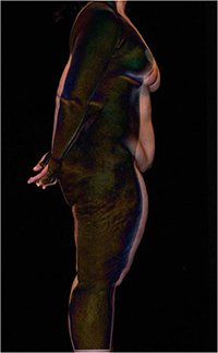
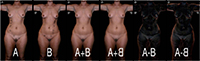
The world of medical imaging exploded into prominence in the late 20th Century. Magnetic Resonance, 3-D CAT scans, and functional PET scans all made it unnecessary to actually see the body. Clinical photography lingered in the analog realm in the 1980s while novel computer-based technologies grabbed scientific and public attention. As these changes occurred, simple patient and disease-related photography slipped in recognition to the level of common and/or old-fashioned. With little remaining momentum for standardization or quantification, medical photographs became devalued, often being characterized as mere visual anecdotes. By the time digital photographic density started to match emulsions accuracy in the 1990s, few in medicine were using clinical images as quantifiable sources for scientific data, and countless clinicians who had access to point-and-shoot technology were satisfied by medical snapshots which flooded the field. After the publication of John Paul Vetters major text of Biomedical Photography in 1992 (Vetter 1992), far too little attention was directed to the production of standardized clinical photographs, and far too little teaching of standards for anatomic capture has taken place. Even in the world of Plastic Surgery, where it is generally acknowledged that anatomically accurate images be produced for every surgical case, photographs are still captured anecdotally. Perhaps more important, there is little published dialog on image quality, no agreement about standards of technique, and no practical or investigative guide available for the scientific analysis of clinical images that are produced. In this new millennium, image analysis software is commonly available for useful investigation of visual information obtained in the visible light spectrum. Although no one would ethically advocate digital alteration of data included in scientific images, non-destructive digital analysis is a valid and underutilized tool for investigation. With urgently needed guidance from biomedical imaging professionals, I would predict that photography will resurface in the world of medicine, but in a more scientifically valid format to answer complex anatomic questions. If standards are articulated for the capture of body images, if rotational techniques are employed (analogous to CAT-scan and MRI methods), and if non-destructive digital analysis is consistently applied, then valuable new data could be reliably and repeatedly obtained. I have used digital analysis of standardized photographs to demonstrate a level of concordance in facial anatomy of monozygotic twins that was never previously recognized (Teplica and Keith 1996). In the last few years alone, commonly available image analysis software was used to define and characterize the twin Mirror Phenomenon which previously had only been rumored to exist without any scientific validation (Teplica and Peekna 2005). In addition, these techniques have been used to begin to answer questions about human embryology and early development (Teplica et al 2007). Finally, the same techniques have preliminarily confirmed mirroring of body form in twin torsos (Figure 1), providing insight on the genetic predetermination of body shape, and explaining the futility of diet and exercise to do anything but temporarily change ones size (Teplica 2009). In each case, standardized visible-spectrum photographs served as the vehicle for data acquisition and analysis. Similarly, the progression of almost any disease or anatomic change could be accurately and repeatedly captured and measured. As an example from the field of Plastic Surgery, if strict imaging standards could also be applied to the often woefully vernacular before and after sequences, then accurate assessment of results could be made (Figure 2) and the quantification of surgical outcomes might soon become routine (Figure 3). It is clear that the possible avenues of photographic investigation are numerous and that their exploration is long overdue. I believe a rich and scientifically valuable future is possible for biomedical images that are quantifiable. Highly standardized photographs harvested with a rotational technique and studied with image-analysis software actually become functional, and can provide a trove of valid data to answer long-held questions about human anatomy, its origins, or its change over time. It is clear that biomedical imaging professionals who embrace both digital technology and standardization must guide the clinical world toward meaningful data acquisition from the visible light spectrum. Presented in part as the Keynote Address to the BioCommunications Association, at BIOCOMM 2009, in Park City, Utah, on 27 July 2009, and as the Keynote Address to the 1st World Congress on Twin Pregnancy: A Global Perspective, in the Palace of San Clemente, Venice, Italy, 16 April 2009. References Teplica, D. Iconography in Twins: A Modern Photographic Perspective, presented as the Keynote Address to the 1st World Congress of Twin Pregnancy: A Global Perspective, Palace of San Clemente, Venice, April 2009; illustrated in larger-than-life full body prints in an accompanying exhibition entitled Twin Shape: Mirroring of Body Form, on the Island of San Servolo, Venice; printed in abstract form as, Teplica, D. Iconography of Twins: A Modern Visual Perspective, Journal of Maternal-Fetal & Neonatal Medicine, Vol. 22, Supp 1, p. 35, March 2009. Teplica, D., and Keith, D., A Study of the Mirror Symmetry Phenomenon in the Faces of 100 Sets of Monozygotic Twins, using Rigidly Standardized Photographic Techniques and Digital Analysis, 10th International Workshop on Multiple Pregnancy, Zakopane, Poland, Archives of Perinatal Medicine, Scientific Publishers OWN, Poznan, Poland, 1996. Teplica, D. and Peekna, K. The Mirror Phenomenon in Monozygotic Twins. In Blickstein, I., and Keith, L., Multiple Pregnancy. 2nd ed. London: Taylor and Francis, 2005, Chapter 38: 277-288. Teplica, D., Derom, C., Peekna, K., and Derom, R., Embryological Timing in Mirror-Image Twinning Twin Research and Human Genetics, Vol. 10, Supplement, p 54, June 2007. Vetter, J. P., ed. Biomedical Photography. Stoneham, MA: Focal Press, 1992. David Teplica is Clinical Associate, Section of Plastic & Reconstructive Surgery Department of Surgery The Pritzker School of Medicine, The University of Chicago; and Attending Surgeon, Saint Joseph Hospital, Chicago, IL. His photography has been widely reproduced, the images are exhibited worldwide, and prints are held in many museum, corporate, and private collections. Please direct all correspondance to: doctor@davidteplica.com |
|
Copyright
2009, The Journal of Biocommunication, All Rights Reserved |

 ]
]