 |
||||
Jan Wandelaar, Bernard Siegfried Albinus and an Indian rhinoceros named Clara set high standards as the process of anatomical illustration entered a new phase of precision, artistic beauty, and marketing in the 18th century |
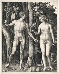
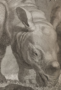
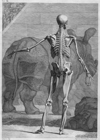
| Linda Wilson-Pauwels The copperplates in Tabulae sceleti et musculorum corporis humani prepared by Jan Wandelaar for Bernard Siegfried Albinus established new production standards in anatomical illustration in the 18th century. The most famous engraving in the atlas depicts a fourth-order muscle man standing in front of the first anatomically correct depiction of a living rhinoceros to arrive in Europe. Wandelaar and Albinus immediately proclaimed the image as a symbol of their book, and engravings of Clara, the rhinoceros, were in the shops in Leiden five years before the book was published in 1747. The engraving acted as an advertisement for both the book and Clara’s pending grand tour of Europe.
|
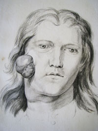
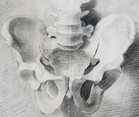
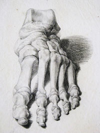
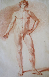
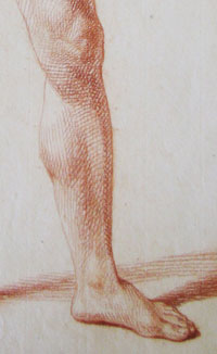
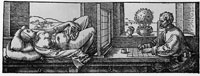
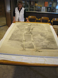
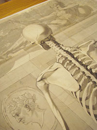
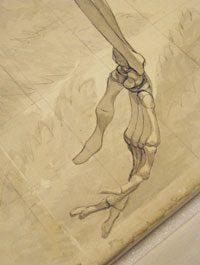
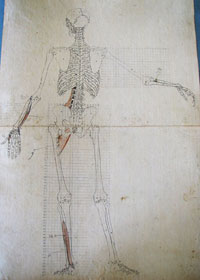
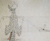
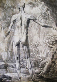
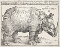
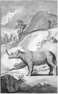
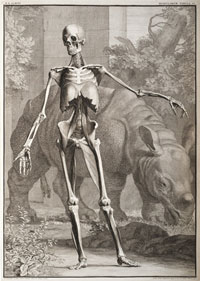
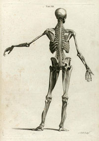
Introduction In the 1700s, the geographical centre for anatomical research and teaching shifted from the Italian schools to universities in the Netherlands (Cazort 1996). During this period, Jan Wandelaar (1690–1759), artist, and Bernhard Siegfried Albinus (1697–1770), chair of Anatomy and Surgery, formed a unique and highly successful artist-anatomist partnership at the University of Leiden. The transfer of knowledge between the two men flourished during a period of Enlightenment ideals, the availability of cadavers for dissection and comparison, and high-quality copperplate engraving. Albinus and Wandelaar first met when they collaborated on a new edition of Andreas Vesalius’s work with Herman Boerhaave (1668–1738), a distinguished professor of botany, chemistry, and medicine. Albinus, as Boerhaave’s student, had worked for many years with him on the adaptation of the classical writings of the Flemish anatomist. In 1723, at the age of 33, Wandelaar joined them in this work. He reproduced the original plates in Opera omnia anatomica & chirugica (1725), Historia musculorum corporis hominis (1734), and Icones ossium foetus humani (1737). After this successful collaboration, Albinus and Wandelaar decided to work together on the great anatomical atlas Tabulae sceleti musculorum corporis humani, (1747)1, arguably the most important anatomical work of the 18th century. A fine collection of sketches of Wandelaar’s work can be found in boxes and crates in the Special Collections Reading Room of the Universiteitsbibliotheek Leiden2. The University of Leiden’s collection of preparatory studies for Tabulae sceleti is unique in the history of anatomical illustration, given its completeness, the importance of the publication, and the extraordinary beauty of the drawings (Cazort et al. 1996). Wandelaar was an exceptional draftsman and copperplate engraver; Albinus had an unrivalled gift for preparing tissue. Artists and anatomists continue to be attracted to their book Tabulae sceleti, as the images are well known for their anatomical precision, beautiful backgrounds, and impressive size. Unlike most artists of the period who drew the anatomical specimens and hired others to engrave the final artwork, Wandelaar engraved all 40 plates, as the signature on each attests and as Albinus states in the preface to the book. The three engravings of an entire skeleton and nine engravings of entire muscle men were produced using a unique illustrative process. The anatomical figures are also set against elaborately drawn landscape backgrounds. The production method and the inclusion of a rhinoceros behind the fourth-order muscle man make this historical publication intriguing and worth further consideration. The Collaboration Between Wandelaar and Albinus Jan Wandelaar Unlike Andreas Vesalius’s anonymous artists, the most renowned of whom is suspected to be Joannes Stephanus of Calcar, Wandelaar’s identity as Albinus’s artist was clear and his life is well documented. Wandelaar was already an accomplished scientific artist before he collaborated with Albinus in 1723. He was trained by Jacob Folkema, the Dutch printmaker; Gillem van der Gouwen, the Dutch engraver and mapmaker; and Gérard de Lairesse, the well-known anatomical artist who produced illustrations for Govert Bidloo’s Anatomia Humani Corporis. Wandelaar had also drawn several anatomical specimens for Fredrik Ruysch, a Dutch botanist and anatomist and planned to produce a book on the human body with his friend and physician, Arend Cant who unfortunately died suddenly before their plans were realized (Punt 1983). Wandelaar’s dramatic use of light and shadow, perspective, contour and form lines demonstrate his fine draftsmanship and competency as an artist and engraver (Figures 1-3). Even in a sketch created with a piece of chalk, the preliminary work of an exceptional artist who thinks like an engraver is clearly evident (Figures 4a & 4b). Bernard Siegfried Albinus For 49 years, Albinus devoted himself to the study of anatomy. “No major anatomist has applied himself so fully to anatomical illustration over so long a period as did Albinus” (Roberts and Tomlinson 1992, p. 322). Albinus was passionate about his work and put it above his personal life as it is documented that he spent 24,000 florins of his own money on the production of the Tabulae sceleti, his 6th book, far more than the 15,000 florins he paid for his house in Leiden (Roberts and Tomlinson 1992; Thornton and Reeves 1983). Born in Frankfurt an der Oder in 1697, Albinus entered the University of Leiden when he was 12 years old. There he frequented the lectures of Govert Bidloo, Johannes Jacobus Rau, Fredericus Dekkers, and Herman Boerhaave. In 1721, when Albinus was 24, he succeeded his father as professor of Anatomy and Surgery at the university. Albinus was greatly influenced by Boerhaave’s mechanistic understanding of the body and attempts to explain its functions by mechanical, hydraulic, and mathematical laws (Hildebrand 2005). He translated this perspective to his own studies of human anatomy and physiology. Didactically, he wanted his students to understand that all human beings are variants of this “homo perfectus” (Punt 1983). In the preface of his classic work of 1747, Albinus wrote, To reproduce, not according to the view, as is customary, but from actual measure, to reproduce what the best in nature displays: to reproduce, not as the demonstrators of anatomy generally do, by merely placing before the eyes of the artist what they have uncovered, but by collecting data from one body after another, and making a composite according to rule so that the actual truth will be displayed, etc. A Mutual Dependence The intense collaboration between Wandelaar and Albinus lasted from 1723, when the two started to work together, until Wandelaar’s death in 1759. This mutually dependent relationship, in which Wandelaar functioned as the creator of the images and Albinus, as their evaluator, created a democratic, if not somewhat forced, union (Punt 1983). It is documented in the literature that Albinus controlled and scrupulously supervised Wandelaar’s work and that he was a difficult task master ordering Wandelaar to redo his illustrations over and over again. The rules that Albinus insisted that Wandelaar follow when creating his illustrations were based on Albinus’s pursuit of the triple symbiosis of objectivity, symmetry, and vitality in anatomical illustration (Hildebrand 2005; Punt 1983). Objectivity referred to the precise depiction of the form and location of the parts of the body, and their relation to other body structures. Symmetry stemmed from the classical Vitruvian sense—aesthetically beautiful with ideal proportions. Vitality equated to strength, beauty, and grace of movement. The figures in Tabulae sceleti represent man, “In control of his world and confident of his status in the Age of Enlightenment, he is truly Homo perfectus” (Cazort et al. 1996, p. 199). After the death of his son, Wandelaar lived in Albinus’ house from about 1746 until he died in 1759. This close working relationship undoubtedly contributed to the accuracy and productivity of Tabulae sceleti. Roberts and Tomlinson wrote (1992) that when Wandelaar died, Albinus was reported to have suffered serious mental depression that he recovered from very slowly. Albinus married six years after Wandelaar’s death and, when he died, his widow sold his library and preserved specimens to the University of Leiden (Cazort et al. 1996). The Process Unlike Vesalius’s work that was produced in 3 years, it seems that Albinus and Wandelaar took 20 years to produce Tabulae sceleti: 13 years to produce the artwork, and possibly 7 to 8 more to engrave it (Cazort et al. 1996; Roberts and Tomlinson 1992). Albinus was critical of Vesalius’ and Bidloo’s atlases for being too pictorial (Cazort et al. 1996). In an attempt to increase the scientific accuracy of anatomical illustration, Albinus and Wandelaar devised a new drawing technique. They placed two nets with square webbing at specified intervals between the artist and the anatomical specimen and copied the images using the grid patterns. This was not an entirely new method. In 1525, Albrecht Dürer used a foramen fixum to fix his viewpoint in his woodcut that illustrated a perspective machine (Figure 5). With this method, the artist depicts each part of the skeleton at a right angle, thus eliminating perspective-induced foreshortening. It was actually the incredible distance between the artist and specimen that made Wandelaar’s and Albinus’s technique novel. To use parallel projection without distorting perspective, Wandelaar drew the cadavers 40 Rhenish feet away from the drawing easel (12.6 m, or 41.2 ft). To improve proportions for scientific accuracy, Wandelaar produced the drawings or washes in 3 phases (see descriptions in Punt 1983, p.18–53, and Roberts and Tomlinson 1992, p. 323–4). First drawing phase The first grid, made of large squares (7.3 x 7.3 cm), was placed directly in front of the cadaver. Wandelaar drew the same pattern of squares on his sheet of paper, looked through a peep-hole (a foramen fixum), using the position of the heart for his sight line, and then broadly sketched the skeleton with a brush and grey wash (Figures 6a, 6b, 6c), producing an image that lacked detail. This process must have been very limiting for an artist of Wandelaar’s abilities. Second drawing phase To achieve more detail, Wandelaar moved his easel to within 4 Rhenish feet (1.3 m, or 4.1 ft) of the cadaver and erected a second grid with 10 times more squares than the first (7.3 x 7.3 mm). He lined up the smaller squares with the larger ones and was able to accurately draw the details (Figures 7a & 7b). To avoid distortion, he picked sight points, such as the hand, that were always at right angles to the part of the body he was drawing. He drew bones using compasses and rulers, and tested for symmetry as a symbol of perfection (Punt 1983). Wandelaar then reduced the skeleton figure onto folio-sized paper marked with a grid of 2.5 cm squares, each of which corresponded to the 7.3 cm squares in the large drawing (Cazort et al. 1996; Punt 1983). Third ‘adaptation’ phase During the process, Albinus and Wandelaar used a collection of different bodies. As a result, they were faced with adapting muscles from different cadavers to fit the original drawings of the skeleton. They created an adaptation formula to reduce or enlarge the muscles until they could be applied to the sketches of the skeleton exactly between the average of the ideal points of origin and insertion (Figure 7a & 7b). Backgrounds The figure-background relationship in the Tabulae sceleti ensures that artists appreciate the publication as much as anatomists. Although Albinus was strict about depicting the human body accurately, he allowed Wandelaar to place his figures in elaborate natural settings (Figure 8), a return to the Vesalian tradition of the anatomical figure-in-a-landscape. Wandelaar suggested the natural backgrounds of the full figure plates in the Tabulae sceleti “in order to preserve the proper light of the picture, for if the space around the figure and between its parts were white, the light would suffer” (Elkins 1986, p. 94). The backgrounds were also intended to relieve the harshness of the figures by providing the illusion of three-dimensionality and to improve the total balance of the figures themselves (Roberts and Tomlinson 1992). But why was an animal included in the background of the fourth-order muscle man? Perhaps Wandelaar was inspired by Albrecht Dürer’s woodcut The Fall of Man (Figure 9) where Dürer positioned the elk behind his ideally proportioned male and female forms. Or perhaps Wandelaar was excited that Clara, the first live rhinoceros to come to Europe, had arrived in Leiden. Clara The most famous plate in Albinus and Wandelaar’s seminal atlas depicts a skeletal male figure standing in front of Clara, an enormous grazing one-horned Asian rhinoceros calf from India (Rhinoceros unicornis Linnaeus, 1758). Until this time, the most copied image of a rhinoceros was Dürer’s woodcut of 1515 (Figure 10), which he based on written descriptions of the animal (Rookmaaker 2005; Ridley 2004). For about the next two hundred years, artists reproduced variations on his engraving showing the species with the fictitious dorsal horn. “Probably no animal picture has exerted such a profound influence on the arts” (Clark 1986, p. 20). Clara was only months old when her mother was killed and she was captured in the Kingdom of Assam, whose ruler presented her to J.A. Sichterman, the director of the Dutch East India Company in Bengal. Sichterman raised Clara as a pet in his home until she became a liability in the dining room. He then sold her to a 36-year-old Dutch sea captain, Douwe Mont van der Meer, who while “…dreaming of a new enterprising future” (Rookmaaker and Monson 2000, p. 315), transported the calf on the ship Knappenhof from India to Rotterdam, Holland, arriving on the 22nd of July, 1741. There Clara started her 16-year grand tour of Europe. Thanks to van der Meer’s flare for public relations (Clarke, 1986), Clara was the center of a European rhinoceros-craze, attracting the attention of many thousands of people, including royalty until she died at the age of 20. She was transported through Europe in a carefully constructed cart that was able to carry her enormous weight of three tons. Her itinerary took into account time to recover from the stress levels of travel and her hormonal cycle. High-quality memorabilia was created in her honour and van der Meer effectively established his copyright over these images (Ridley 2004). When Albinus and Wandelaar first saw Clara in 1741 in the Amsterdam Zoo, they immediately proclaimed her to be a symbol for their atlas (Punt 1983). Perhaps this declaration stemmed from Wandelaar’s excitement as this was not the first time that he had drawn and engraved a rhinoceros. He engraved a two-horned black African rhinoceros (Diceros bicornis Linnaeus, 1758) for the Dutch translation (1727) of Peter Kolb’s Caput Bonae Spei Hodiernum, originally published in German in 1719 (Rookmaaker 1976). During the task of copying some of the plates of the German edition for the Dutch translation, Wandelaar adapted his engraving to match Kolb’s description of the black rhinoceros (Figure 11). Later he received advice from James Douglas3 who, in letters found in his bound volume of rhinoceros drawings and engravings dated 17394, recommended that Wandelaar make corrections to the head, and fore and hind feet (see Wandelaar’s sketches dated 1734–35, Plate 2, in Rookmaaker 1976). All of this happened many years before Wandelaar drew Clara. His excitement when he had the opportunity to draw the first living rhinoceros calf on European soil can only be imagined. The most famous engraving of the fourth-order muscle man stands out prominently against a background that includes Clara (Figure 12a & b). There is no doubt that Wandelaar’s fine copper engraving of Clara is drawn from life because it depicts, in three-quarters view, a young calf with an undeveloped horn, grazing leisurely. Wandelaar was recognized as the first to record and publish Clara’s likeness when she was a 2½- to 3-year-old calf in 1741. Clarke (1986) writes, “It is the earliest life-like image by an artist of talent” (p. 193). Many other posters printed from woodcuts and engravings, dated from 1746 to 1756, were made of Clara during her tour of Europe. The production dates can be judged from Clara’s age, based on her weight and the development of her horn (see list in Rookmaker and Monson 2000, Table 1). Wandelaar’s engraving of Clara was put on sale separately from Tabulae sceleti (Ridley 2004) and loose plates of the engraving were available in the shops of Leiden in 1742 (Riley 2004; Clark 1986), one year after Clara’s arrival in Leiden and five years before the Latin edition of Albinus’s Tabulae sceleti was available. Appearing singly in this way, Wandelaar’s image could be seen as an accomplished precursor of a twentieth-century marketing campaign (Ridley 2004). The engraving therefore served a dual purpose, to advertise Clara’s presence in Leiden and to advertise the forthcoming publication of the new medical book Tabulae sceleti. Conclusion It took 8 years to engrave the 40 large copperplates in Tabulae sceleti and not a single image was produced freehand. Although Albinus did not give Wandelaar the credit that he deserved for his contribution to Tabulae sceleti, Elkin (1986) wrote that Albinus did marvel at Wandelaar’s preparatory sketches and that Albinus remarked that artists were astonished that Wandelaar could have made such elaborate engravings from such sketches. Their work in Tabulae sceleti influenced generations of anatomists and artists. “On the facing page of each finished etching, Wandelaar and Albinus included a print of the original line etching keyed to the text, thus eliminating the confusion of letters or numbers on the main print. This innovation was adopted by anatomical illustrators well into the nineteenth century” (Cazort et al. 1996, p. 199). Although when published, the Tabulae sceleti was widely criticized for its background motifs (Hildebrand 2005, Elkins1986), Wandelaar justified his use of unusual backgrounds in the 1749 reprinted English edition of Tabulae sceleti re-engraved by Charles Grignion (Figure 13).Wandelaar wrote about the two engravings including images of Clara, “We thought the rarity of the beast would render these figures of it more agreeable than any other ornament, resulting from mere fancy. The figures are just, and of magnitude proportionable to the human figures contained in theses two tables” (Clark 1986). Even after criticism, the images were copied in many anatomical books, however, in the 1777–1778 Edinburgh publication, re-engraved by the famous Andrew Bell, the elaborate backgrounds were removed for clarity (Figure 14). Although Albinus writes in the preface of the book, “And thus he [Wandelaar] was instructed, directed, and as entirely ruled by me, as if he was a tool in my hands, and I made the figures myself,” the exceptional collection of sketches by Wandelaar at the University of Leiden attests to the fact that Wandelaar was a very competent artist. Not only did Wandelaar’s copper engravings with elaborate backgrounds, including the famous rhinoceros Clara, ensure the success of the atlas, they assured Wandelaar an eminent place amongst anatomical artists and anatomists.
References Cazort M., M. Kornell, and K.B. Roberts.1996. The ingenious machine of nature: Four centuries of art and anatomy. Ottawa: National Gallery of Canada Publisher. Choulant. L. 1920. History and bibliography of anatomic illustration. Translated and annotated by Mortimer Frank. Chicago: University of Chicago Press. Clarke, T. H. 1986. The rhinoceros from Dürer to Stubbs: 1515–1799. London: Sotheby's Publications. Hildebrand, R. 2005. Attic perfection in anatomy: Bernhard Siegfried Albinus (1697-1770) and Samuel Thomas Soemmerring (1755-1830). Annals of Anatomy 187:555-573. Kemp, M., and M. Wallace. 2000. Spectacular bodies: The art and science of the human body from Leonardo to now. Published jointly by the Hayward Gallery and the University of California Press, London. Punt, H. 1983. Bernard Siegfried Albinus (1697-1770) ‘On human nature’ anatomical and physiological ideas in eighteenth century Leiden. Amsterdam: B. M. Israel. Ridley, G. 2004. Clara’s Grand Tour: Travels with a rhinoceros in eighteenth-century Europe. New York: Atlantic Monthly Press. Roberts, K.B., and J.D.W. Tomlinson. 1992. The fabric of the body. Oxford: Oxford University Press. Rookmaaker, L.C. and J. Monson. 2000. Woodcuts and engravings illustrating the journey of Clara, the most popular rhinoceros of the eighteenth century. Der Zoologische Garten 70(5):313-335. Rookmaaker L.C. 1976. An early engraving of the black rhinoceros (Diceros bicornis [L]) made by Jan Wandelaar. Biological Journal of the Linnean Society 8:87-90. Sappol M. 2002. Dream Anatomy. Washington: National Library of Medicine. Strieder P. 1982. Albrecht Dürer • Paintings • Prints • Drawings. New York, Abaris Books Inc. Thomas Fisher Rare Book Library, University of Toronto, Anatomia Digital Collection. Available online, http://link.library.utoronto.ca/anatomia/application/index.cfm (accessed August 5, 2008). Thornton, J.L. and C. Reeves. 1983. Medical book illustration. Cambridge: The Orleander Press.
Acknowledgements A special thanks to Drs. Harm Beukers and Andre Bouwman, Universiteitsbibliotheek Leiden, Leiden University. The author photographed Wandelaar's original artwork in the Special Collections Reading Room of the Universiteitsbibliotheek in Leiden (Figures 1, 2, 3, 4a, 4b, 6b, 6c, 7a, 7b, and 8). Figure 6a was photographed by André Bouwman.
|
Copyright
2009, The Journal of Biocommunication, All Rights Reserved
Table
of Contents for VOLUME 35, NUMBER 1