Ophthalmic Photographer’s Society 2007 Awards
The Gallery for this issue of JBC features the award-winning images from the 2007 Ophthalmic Photographers’ Society’s Scientific Exhibit. The purpose of each annual OPS Scientific Exhibit is to encourage members to share their outstanding images with the ophthalmic community. The entire exhibit, featuring 31 different categories in the print and stereo divisions, is displayed as part of the Scientific Exhibit at the Annual Meeting of the American Academy of Ophthalmology.
The Ophthalmic Photographers' Society is a non-profit organization dedicated to a highly specialized form of medical photography pertaining to the field of ophthalmology. The main objectives of the Society are to provide primary and continuing education in the field of ophthalmic photography, to set and maintain standards for the profession through a multi-level certification program, and to promote scientific advancement in the technology.
For additional information about OPS, please visit: http://www.opsweb.org/
Also JBC featured a two part series (in Volume 32 No. 2 and, Volume 33 No. 1) on the topic entitled: Ophthalmic Imaging - An Overview and Current State of the Art.
| Print Division |
| |
| Best of Show |
| |
|
| |
| First Place |
| |
|
| |
|
| |
|
| |
|
| |
|
|
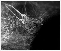
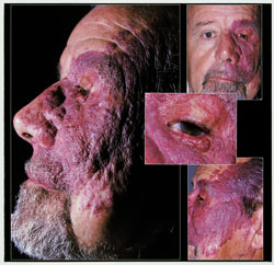
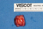
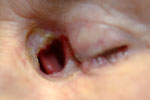
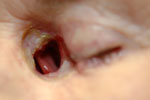
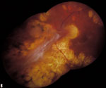
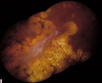
Port Wine Stain
External Photography
Chris Barry, CRA, FOPS ©
Lions Eye Institute, Perth,
Western Australia
|
|
| |
|
|
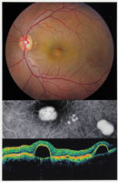
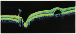
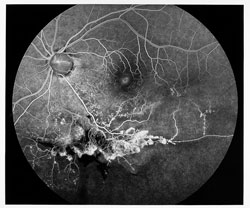
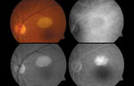
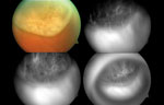
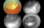
Central Serous Choroidopathy
Matthew Olson, CRA ©
University of Mississippi Medical Center, Jackson, MS
|
|
| |
|
| |
|
| |
|
|
Extreme Laser
Marshall E. Tyler, CRA, FOPS ©
Wake Forest University Eye Center, Winston-Salem, NC
|
|
| |
|
| |
|
| |
| Second Place |
| |
|
|
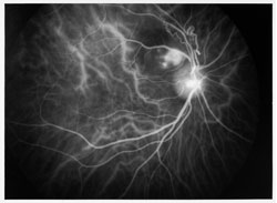
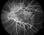
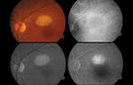
Branch Retinal Vein Occlusion
Matthew Olson, CRA ©
University of Mississippi Medical Center, Jackson, MS
|
|
| |
|
|
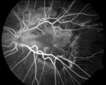
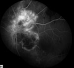
CNV/BRVO
ICG Angiogram
Lisa Schillace, CRA ©
Henry Ford Health System, Detroit, MI
|
|
| |
|
|
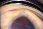
Iridotomy
Gonio Photography
Leslie MacKeen, CRA ©
|
|
| |
|
|
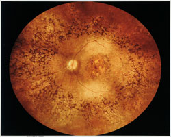
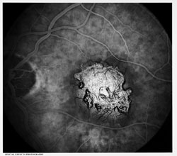
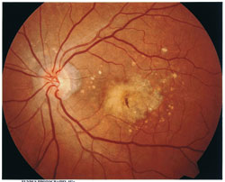
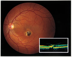
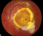
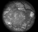
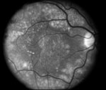
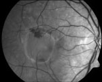
AMD
Fundus Photography 45°+
Denise Armiger, CRA ©
Columbia University, NY
|
|
| |
|
|
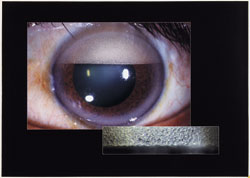
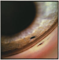
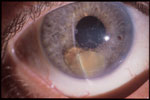
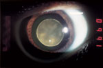
Marfans Syndrome
Slit Lamp Photography
Marriner L. Skelly, CRA ©
Duke University Eye Center, Durham, NC
|
|
| |
|
|
ERM with Scar
Cross Categories
Jarron Wehmeier ©
Barnes Retina Institute, St. Louis, MO
|
|
| |
|
|
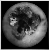
Congenital Hypertrophy of the RPE
Monochromatic Photography
Marriner L. Skelly, CRA ©
Duke University Eye Center, Durham, NC
|
|
| |
|
|
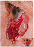
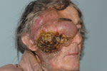
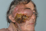
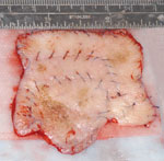
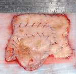
Basel Cell Carcinoma
David Miller, CRA ©
Wake Forest University Eye Center, Winston-Salem, NC
|
|
| |
|
|
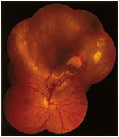
Hemangioblastoma
Matthew Raeber ©
Barnes Retina Institute, St. Loius, MO
|
|
| |
|
|
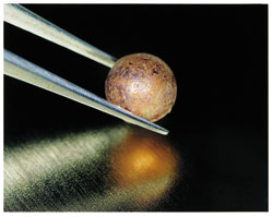
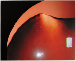
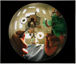
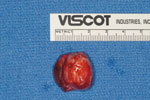
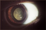
IOL Implantation (From a Patient’s View)
Chris Barry, CRA, FOPS ©
Lions Eye Institute, Perth,
Western Australia
|
|
| |
|
|
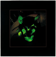
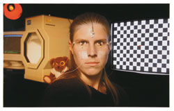
Mouse After Fluorescein
Special Effects Photography
Leslie MacKeen, CRA ©
|
|
| |
|
|
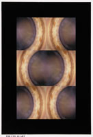
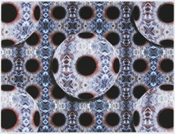
Iris Mozaic
The Eye as Art
James Gilman, CRA ©
Moran Eye Center, Salt Lake City, UT
|
|
| |
|
|
Electroretinogram
Chris Barry, CRA, FOPS ©
Lions Eye Institute, Perth, W. Australia
|
|
| |
| |
| Stereo Slide Division |
| |
| Best of Show |
| |
|
|
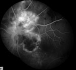
Combined Hartoma of the Retina and Retinal Pigment Epithelium
Fluorescein Angiogram
Pablo Gili, MD ©
Fundacion Hospital, Alcorcon, Spain
|
|
| |
| First Place |
| |
|
|
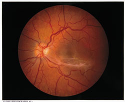
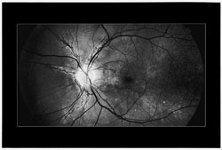
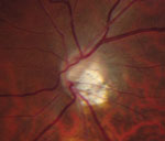
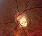
Glaucoma
Fundus Photography 20°
Robert Prusak, CRA ©
Kellogg Eye Center, Ann Arbor, MI
|
|
| |
|
|
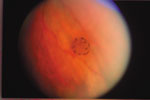
Untitled
Fundus Photography 30°–40°
Charles Hamm, CRA, COT ©
Mason Eye Institute, Columbia, MO
|
|
| |
|
|
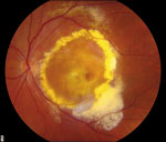
ARMD with Fibrosis Macular Scar
Fundus Photography 45°+
Jim Sherry, CRA ©
The Macula Center, Henderson, NC
|
|
| |
|
|
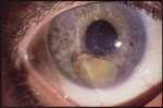
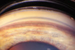
Subluxed Lens in the AC
Sit Lamp Photography
Michael Stanley ©
Medical College of Georgia, Govetown, GA
|
|
| |
|
|
Basal Cell Carcinoma
External Photography
Jay Rostvold ©
Mayo Clinic, Rochester, MN
|
|
| |
|
| |
|
| |
|
|
Macular Dystrophy
Monochromatic Photography
|
|
| |
|
|
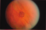
Untitled
Composite
Christina Malik Consolo, CRA, COT
Associated Retina Consultants, Rochester, MI
|
|
| |
|
|
Retinal Astrocytic Hamartoma
Cross Categories
Pablo Gili, MD
Fundacion Hospital, Alcorcon, Spain
|
|
| |
| Second Place |
| |
|
| |
|
|
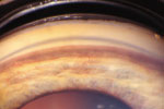
Untitled
Slit Lamp Photography
Zvi Friedman ©
Sheba Medical Center, Ramatgan, Isreal
|
|
| |
|
|
Basal Cell Carcinoma
External Photography
Sarah Moyer, CRA ©
Durham, NC
|
|
| |
|
|
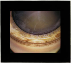
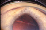
Ciliary Body Melanoma with Hemorrhage
Gonio Photography
Jay Rostvold ©
Mayo Clinic, Rochester, MN
|
|
| |
|
|
PED
Monochromatic Photography
Pablo Gili, MD ©
Fundacion Hospital, Alcorcon, Spain
|
|
| |
|
| |
|
|
Choroidal Melanoma
Cross Categories
Pablo Gili, MD ©
Fundacion Hospital, Alcorcon, Spain
|
|
| |
| |
|
|